. 2022 Jul 14;79(8):420.
doi: 10.1007/s00018-022-04448-2.

Molecular crowding facilitates bundling of IMPDH polymers and cytoophidium formation

Affiliations

Molecular crowding facilitates bundling of IMPDH polymers and cytoophidium formation

Chia-Chun Chang et al. Cell Mol Life Sci. .

Abstract

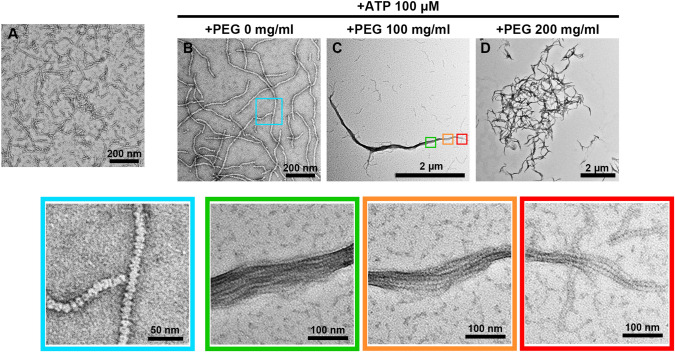
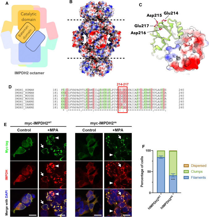
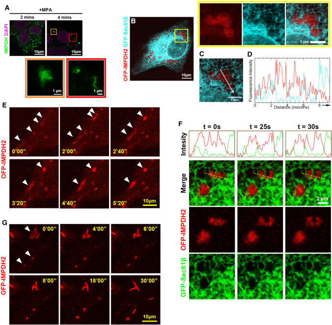
The cytoophidium is a unique type of membraneless compartment comprising of filamentous protein polymers. Inosine monophosphate dehydrogenase (IMPDH) catalyzes the rate-limiting step of de novo GTP biosynthesis and plays critical roles in active cell metabolism. However, the molecular regulation of cytoophidium formation is poorly understood. Here we show that human IMPDH2 polymers bundle up to form cytoophidium-like aggregates in vitro when macromolecular crowders are present. The self-association of IMPDH polymers is suggested to rely on electrostatic interactions. In cells, the increase of molecular crowding with hyperosmotic medium induces cytoophidia, while the decrease of that by the inhibition of RNA synthesis perturbs cytoophidium assembly. In addition to IMPDH, CTPS and PRPS cytoophidium could be also induced by hyperosmolality, suggesting a universal phenomenon of cytoophidium-forming proteins. Finally, our results indicate that the cytoophidium can prolong the half-life of IMPDH, which is proposed to be one of conserved functions of this subcellular compartment.

Keywords: Cellular compartmentalization; Cytoophidium; IMPDH; Membraneless organelle; Molecular crowding.


Conflict of interest statement

The authors have no relevant financial or non-financial interests to disclose.

Figures

Fig. 1
Fig. 1
Molecular crowder PEG-4000 promotes in vitro reconstitution of IMPDH cytoophidium. AD Negative staining images of hIMPDH2 recombinant protein incubated with different conditions for 1 h. A hIMPDH2 protein was incubated in the buffer without the supplementation of ligands. BD hIMPDH2 protein was incubated in the buffer containing 100 μM ATP and PEG-4000 at 0, 100, 200 mg/ml. Lower panels show magnified images of corresponding areas in B and C
Fig. 2
Fig. 2
The mutation at loop214−217 of human IMPDH2 disturbs the cytoophidium assembly in cells. A Illustration of the IMPDH octamer. B Electrostatic surface model of hIMPDH2 polymer. C Electrostatic surface model of the Bateman domain of hIMPDH2. Residues at loop214−217 are indicated. D The sequence comparison of the CBS subdomain of human, mouse and zebrafish IMPDH isoforms. Residues with positive and negative charge are highlighted in green and red, respectively. E Immunofluorescence of HEK 293T cells overexpressing myc-hIMPDH2WT and myc-hIMPDH24A. Cells were treated with MPA for 2 h before fixation. Cells displaying IMPDH filaments are indicated with arrows and cells displaying IMPDH clumps are indicated with arrowheads. Scale bars = 20 μm. F Quantification of cells displaying different IMPDH patterns in (E). 3 biological repeats, total 416 and 367 cells were counted in myc-hIMPDH2WT and myc-hIMPDH24A, respectively. Error bars = S.E.M. Student’s t test, ***p < 0.001
Fig. 3
Fig. 3
Amorphous IMPDH clump is the precursor state of the filamentous cytoophidium. A Immunofluorescence of wild-type HeLa cells treated with MPA for 2 and 4 min, respectively. Magnified images of an amorphous IMPDH clump (orange box) and filamentous cytoophidia (red box) are corresponding to selected areas in A. B Images of HeLa cells expressing OFP-IMPDH2 and GFP-Sec61β fusion proteins. Cells were treated with MPA for 1 h. Magnified images show an amorphous IMPDH clump surrounded by the ER in selected areas. C Single plane image of the IMPDH clump shown in magnified images in B. D Fluorescent intensity of OFP-IMPDH2 and GFP-Sec61β signals in C. The x axis corresponds to the direction and area of measurement indicated by the arrow in C. EG Representative frames of live-cell imaging of OFP-IMPDH2 expressing HeLa cells treated with MPA. E Selected frames of movie S1 showing the movement and fusion of IMPDH clumps (arrowheads). F Selected frames of movie S2 showing an IMPDH clump split in association with ER tubule dynamics. The dashed line corresponds to the area of fluorescence intensity measurement. G Selected frames of movie S3 showing the clump-to-filament transition. Time intervals of each frame is 40 s in E and 60 s in G
Fig. 4
Fig. 4
Hyperosmotic medium induces IMPDH cytoophidium assembly in multiple cell lines. A Immunofluorescence of wild-type HEK 293T cells treated with sucrose at different concentrations for 1 h. B Quantification of the percentage of cells with cytoophidia under the treatments shown in A. 3 biological repeats, total 418, 373, 384, 362 and 470 cells were counted in each group. C Immunofluorescence of wild-type HeLa, MCF7 and HCT116 cells treated with sucrose at different concentrations for 1 h. D Quantification of the percentage of cells with cytoophidia under the treatments shown in C. 3 biological repeats, total 753, 483, 574, and 497 cells were counted in each HeLa cell group. Total 506, 563, 608, and 259 cells were counted in each MCF7 cell group. Total 624, 647, 373, and 570 cells were counted in each HCT116 cell group. Scale bars = 20 μm in all panels. Error bars = S.E.M. Tukey’s test was used in the comparison in B and D
Fig. 5
Fig. 5
Hyperosmotic medium induces the protein polymerization and aggregation of filamentous polymers. A Immunofluorescence of wild-type HEK 293T cells treated with 300 mM sucrose for different periods of time. B Immunofluorescence of wild-type HEK 293T cells pre-treated with 300 mM sucrose for 3 h then treated with isosmotic medium for 1 to 5 min. C and D Immunofluorescence of wild-type HEK 293T cells pre-treated with 100 μM guanosine for 1 h and then treated with 100 μM MPA (C) or sucrose (D). E Quantitative data of the presence of hyperosmolality-induced cytoophidia in HEK 293T cells pre-treated with (+ Gua) and without guanosine (− Gua). 3 biological repeats, total 373, 384, 362 and 470 cells were counted in each − Gua group. 3 biological repeats, total 552, 572, 568, and 583 cells were counted in each + Gua group. (F) Immunofluorescence of wild-type HEK 293T cells for hyperosmolality-induced CTPS, PRPS and IMPDH cytoophidium. G Immunofluorescence of wild-type HEK 293T cells for CTPS, PRPS and IMPDH cytoophidium in the medium containing 300 mM sorbitol. H and I Immunofluorescence for IMPDH and CTPS in HEK 293T cells expressing OFP-P2A-IMPDH2WT, OFP-P2A-IMPDHY12A, Flag-CTPS1WT and Flag-CTPS1H355A. Proportion shown at left-bottom indicates cytoophidium positive cells/total transfected cells in each group. Scale bars = 20 μm in all panels. Error bars = S.E.M. Student’s t test, **p < 0.01 in (E)
Fig. 6
Fig. 6
Inhibition of transcription perturbs the MPA-induced IMPDH cytoophidium assembly. A Immunofluorescence of wild-type HEK 293T cells treated with DMSO or 1 μM CX-5461 for 3 h prior to 15 and 30 min of MPA treatment. B Immunofluorescence of wild-type HEK 293T cells treated with 1 μM ACTD for 1 h before or after the treatment of MPA. C Quantitative data of the abundance of IMPDH cytoophidia under different treatments shown in A and B. 3 biological repeats, total 1023 and 856 cells were analyzed in + MPA 15 min groups. 4 biological repeats, total 737, 681 and 717 cells were analyzed in + MPA 30 min groups. D Immunofluorescence of wild-type HEK 293T cells treated with ACTD for 1 h and sucrose for another 1 h prior to cytoophidium induction with MPA. E Quantitative data of the proportion of cells with cytoophidia in the conditions shown in D. 3 biological repeats, total 554, 519 and 459 cells were counted in each group. Scale bars = 20 μm in all panels. Error bars = S.E.M. Student’s t test, **p < 0.01, *** p < 0.001, **** p < 0.0001 in C and E
Fig. 7
Fig. 7
Formation of the cytoophidium prolongs the half-life of IMPDH2 protein. A Immunofluorescence of myc-IMPDH2WT and myc-IMPDH2Y12A overexpressing HeLa cells treated with DAU or MPA for 1 day. B Western blotting for the exogenous myc-IMPDH2 levels in transfected HeLa cells treated with DAU or MPA for 1 and 3 days. C Quantitative data of the relative myc-IMPDH2 levels in the western blotting shown in (B). Scale bars = 20 μm in all panels. Error bars = S.E.M. Student’s t test, *p < 0.05
Fig. 8
Fig. 8
Illustration of the proposed molecular mechanism for the assembly of the cytoophidium. 1. Protein oligomers (octamer for IMPDH) polymerize into filamentous polymers under the regulation of conformational changes driven by the binding of ligands. Such conformational changes may expose or hide the interfaces responsible for the interactions between protomers. 2. Polymers self-associate into filament bundles through electrostatic interactions, which could be regulated by salt, pH, post-translational modifications, protein concentrations and molecular crowding. Such compact structures of filament bundles may allow small molecules percolate through the aggregate but restrict the interaction between macromolecules and component proteins. 3. Filament bundles may further assemble into larger structures, such as the cytoophidium

Similar articles

Cited by

References

    1. Liu JL. The cytoophidium and its kind: filamentation and compartmentation of metabolic enzymes. Annu Rev Cell Dev Biol. 2016;32:349–372. - PubMed
    1. Liu JL. Intracellular compartmentation of CTP synthase in Drosophila. J Genet Genom. 2010;37(5):281–296. - PubMed
    1. Zhang B, Tastan OY, Zhou X, Guo CJ, Liu X, Thind A, Hu HH, Zhao S, Liu JL. The proline synthesis enzyme P5CS forms cytoophidia in Drosophila. J Genet Genom. 2020;47(3):131–143. - PubMed
    1. Zhang S, Ding K, Shen QJ, Zhao S, Liu JL. Filamentation of asparagine synthetase in Saccharomyces cerevisiae. PLoS Genet. 2018;14(10):e1007737. - PMC - PubMed
    1. Chang CC, Lin WC, Pai LM, Lee HS, Wu SC, Ding ST, Liu JL, Sung LY. Cytoophidium assembly reflects upregulation of IMPDH activity. J Cell Sci. 2015;128(19):3550–3555. - PMC - PubMed

MeSH terms

LinkOut - more resources