

Antibiotic-Derived Radiotracers for Positron Emission Tomography: Nuclear or "Unclear" Infection Imaging?
- PMID: 35834311
- PMCID: PMC9826354
- DOI: 10.1002/anie.202204955
Antibiotic-Derived Radiotracers for Positron Emission Tomography: Nuclear or "Unclear" Infection Imaging?
Abstract
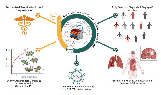
The excellent features of non-invasive molecular imaging, its progressive technology (real-time, whole-body imaging and quantification), and global impact by a growing infrastructure for positron emission tomography (PET) scanners are encouraging prospects to investigate new concepts, which could transform clinical care of complex infectious diseases. Researchers are aiming towards the extension beyond the routinely available radiopharmaceuticals and are looking for more effective tools that interact directly with causative pathogens. We reviewed and critically evaluated (challenges or pitfalls) antibiotic-derived PET radiopharmaceutical development efforts aimed at infection imaging. We considered both radiotracer development for infection imaging and radio-antibiotic PET imaging supplementing other tools for pharmacologic drug characterization; overall, a total of 20 original PET radiotracers derived from eleven approved antibiotics.
Keywords: antibiotic-derived PET; imaging of infection; positron emission tomography; radiolabeling; radiotracers.
© 2022 The Authors. Angewandte Chemie International Edition published by Wiley-VCH GmbH.
Conflict of interest statement
The authors declare no conflict of interest.
Figures
Similar articles
-
(18)F-labeled positron emission tomographic radiopharmaceuticals in oncology: an overview of radiochemistry and mechanisms of tumor localization.Semin Nucl Med. 2007 Nov;37(6):400-19. doi: 10.1053/j.semnuclmed.2007.08.004. Semin Nucl Med. 2007. PMID: 17920348 Review.
-
Update on PET radiopharmaceuticals: life beyond fluorodeoxyglucose.Radiol Clin North Am. 2004 Nov;42(6):1033-53, viii. doi: 10.1016/j.rcl.2004.08.009. Radiol Clin North Am. 2004. PMID: 15488556 Review.
-
Radiopharmaceuticals in positron emission tomography: present situation and future perspectives.Radiologia. 2016 Nov-Dec;58(6):468-480. doi: 10.1016/j.rx.2016.07.003. Epub 2016 Aug 31. Radiologia. 2016. PMID: 27592111 English, Spanish.
-
A Team for Unique Molecular Imaging Based in South Africa.Angew Chem Int Ed Engl. 2023 Aug 1;62(31):e202305726. doi: 10.1002/anie.202305726. Epub 2023 Jun 12. Angew Chem Int Ed Engl. 2023. PMID: 37306614
-
Development of (18)F-labeled radiotracers for neuroreceptor imaging with positron emission tomography.Neurosci Bull. 2014 Oct;30(5):777-811. doi: 10.1007/s12264-014-1460-6. Epub 2014 Aug 29. Neurosci Bull. 2014. PMID: 25172118 Free PMC article. Review.
Cited by
-
Modern tools in cardiac imaging to assess myocardial inflammation and infection.Eur Heart J Open. 2023 Mar 3;3(2):oead019. doi: 10.1093/ehjopen/oead019. eCollection 2023 Mar. Eur Heart J Open. 2023. PMID: 37006410 Free PMC article. Review.
-
PET/CT Imaging of Infectious Diseases: Overview of Novel Radiopharmaceuticals.Diagnostics (Basel). 2024 May 17;14(10):1043. doi: 10.3390/diagnostics14101043. Diagnostics (Basel). 2024. PMID: 38786341 Free PMC article. Review.
-
First-in-human infection imaging with 89Zr-labelled leukocytes and comparison of scan quality with [99mTc]Tc-HMPAO-labelled leukocytes.Front Nucl Med. 2024 Jul 22;4:1426650. doi: 10.3389/fnume.2024.1426650. eCollection 2024. Front Nucl Med. 2024. PMID: 39355210 Free PMC article.
-
Bacteria-targeted imaging using vancomycin-based positron emission tomography tracers can distinguish infection from sterile inflammation.Eur J Nucl Med Mol Imaging. 2025 Apr;52(5):1878-1889. doi: 10.1007/s00259-024-06997-z. Epub 2024 Nov 29. Eur J Nucl Med Mol Imaging. 2025. PMID: 39609275 Free PMC article.
-
Novel Diagnostic Methods for Infective Endocarditis.Int J Mol Sci. 2024 Jan 19;25(2):1245. doi: 10.3390/ijms25021245. Int J Mol Sci. 2024. PMID: 38279244 Free PMC article. Review.
References
-
- Murray C. J. L., Ikuta K. S., Sharara F., Swetschinski L., Aguilar G. R., Gray A., Han C., Bisignano C., Collabora A. R., et al., Lancet 2022, 399, 629–655. - PubMed
-
- Palestro C. J., Semin. Nucl. Med. 2020, 50, 23–34. - PubMed
-
- Pijl J. P., Nienhuis P. H., Kwee T. C., Glaudemans A., Slart R., Gormsen L. C., Semin. Nucl. Med. 2021, 51, 633–645. - PubMed
-
- Goldsmith S. J., Vallabhajosula S., Semin. Nucl. Med. 2009, 39, 2–10. - PubMed
Publication types
MeSH terms
Substances
LinkOut - more resources
Full Text Sources
Medical
