. 2022 Sep 1;132(17):e159876.
doi: 10.1172/JCI159876.

Frequent detection but lack of infectivity of SARS-CoV-2 RNA in presymptomatic, infected blood donor plasma

Affiliations

Frequent detection but lack of infectivity of SARS-CoV-2 RNA in presymptomatic, infected blood donor plasma

Paula Saá et al. J Clin Invest. .

Abstract

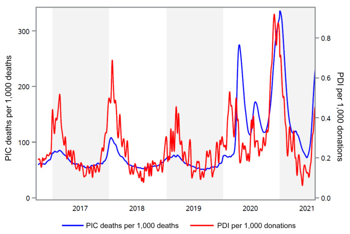
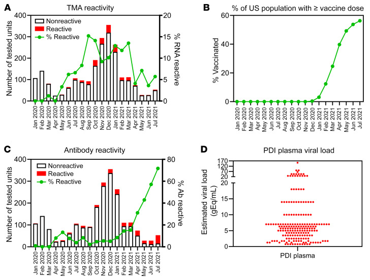
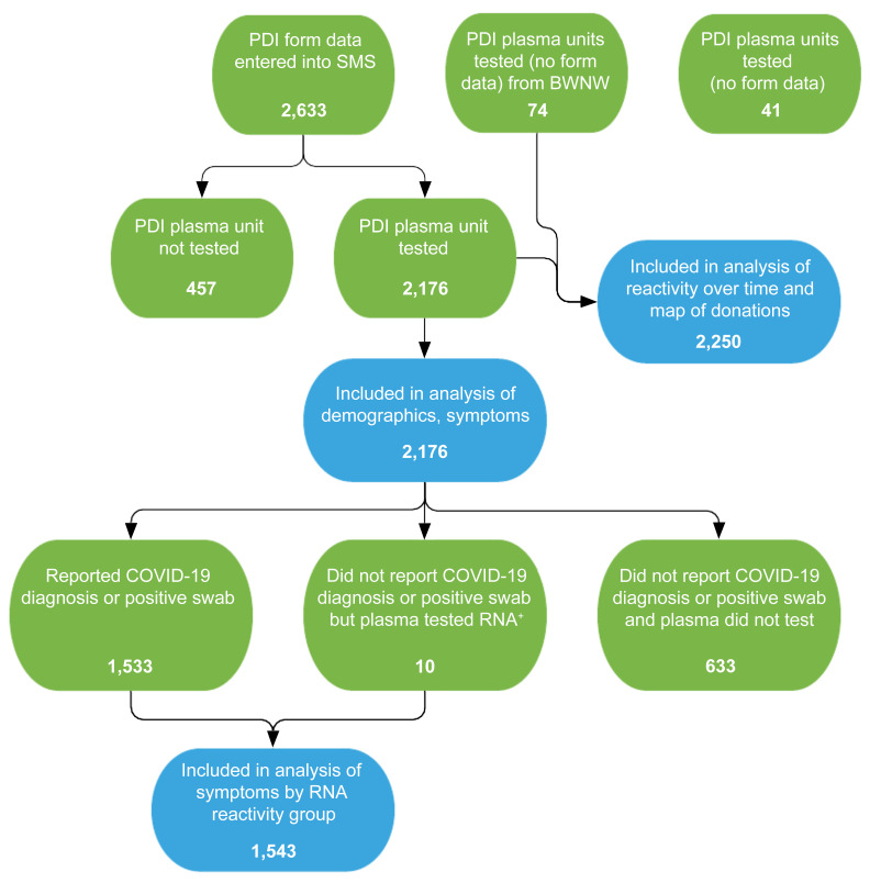
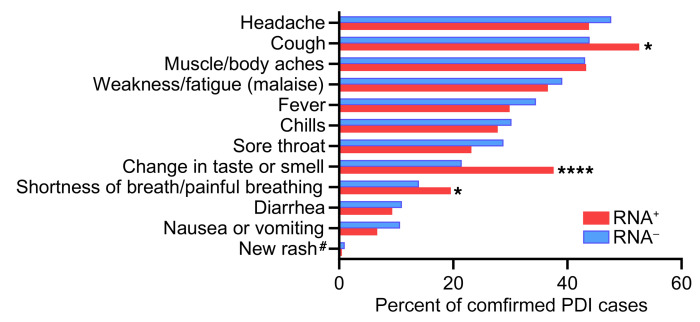
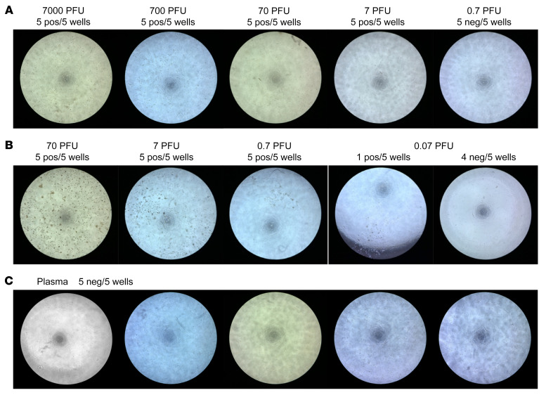
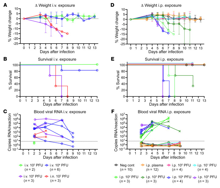
Respiratory viruses such as influenza do not typically cause viremia; however, SARS-CoV-2 has been detected in the blood of COVID-19 patients with mild and severe symptoms. Detection of SARS-CoV-2 in blood raises questions about its role in pathogenesis as well as transfusion safety concerns. Blood donor reports of symptoms or a diagnosis of COVID-19 after donation (post-donation information, PDI) preceded or coincided with increased general population COVID-19 mortality. Plasma samples from 2,250 blood donors who reported possible COVID-19-related PDI were tested for the presence of SARS-CoV-2 RNA. Detection of RNAemia peaked at 9%-15% of PDI donors in late 2020 to early 2021 and fell to approximately 4% after implementation of widespread vaccination in the population. RNAemic donors were 1.2- to 1.4-fold more likely to report cough or shortness of breath and 1.8-fold more likely to report change in taste or smell compared with infected donors without detectable RNAemia. No infectious virus was detected in plasma from RNAemic donors; inoculation of permissive cell lines produced less than 0.7-7 plaque-forming units (PFU)/mL and in susceptible mice less than 100 PFU/mL in RNA-positive plasma based on limits of detection in these models. These findings suggest that blood transfusions are highly unlikely to transmit SARS-CoV-2 infection.

Keywords: COVID-19; Clinical practice; Molecular diagnosis.


Figures

Figure 1
Figure 1. Comparison of the PDI rate with reported mortality due to pneumonia, influenza, and COVID-19.
The red line represents the rate of PDI reports per 1,000 donations to the American Red Cross from week 40, 2016 through week 31, 2021 (right axis). The blue line shows data published by the CDC for the number of deaths from pneumonia, influenza, or COVID-19 per 1,000 deaths (PIC, left axis). CDC data were obtained from the website https://www.cdc.gov/flu/weekly/index.htm
Figure 2
Figure 2. Detection of SARS-CoV-2 RNA and antibodies in PDI donors.
(A) Plasma samples (n = 2,250) were collected over the course of 19 months throughout the United States. Bars correspond to total number of donations tested for SARS-CoV-2 RNA per month of collection, with the red portion denoting those reactive. Line corresponds to percentage of RNA-reactive donations. (B) Data published on the CDC website (https://data.cdc.gov/Vaccinations/COVID-19-Vaccination-Trends-in-the-United-States-N/rh2h-3yt2) show the average proportion of the US population that had received at least 1 vaccine dose for each month of the period during which PDI plasmas were tested. (C) Plasma samples (n = 2,250) were collected over the course of 19 months throughout the United States. Bars correspond to total number of donations tested for SARS-CoV-2 spike antibodies per month of collection, with the red portion denoting those reactive. The green line corresponds to percentage of antibody-reactive donations. (D) Estimated viral load of SARS-CoV-2 RNA in 196 reactive plasma samples based on TMA reactivity in replicates normalized to a standard curve.
Figure 3
Figure 3. Flowchart of PDI donor enrollment and analytic groups.
PDI donor questionnaires were entered into the study management system (SMS). Questionnaires were not obtained from PDI donors at Bloodworks Northwest (BWNW). A total of 2,250 plasma units were available for SARS-CoV-2 RNA testing, and 2,176 had questionnaire data for analysis of demographics and symptoms. Of these donors, 1,543 were determined to be SARS-CoV-2 infected based on a self-report of a positive SARS-CoV-2 clinical test or by detection of RNAemia in the donor’s plasma unit. Green bubbles indicate the parent populations and blue bubbles indicate analysis populations.
Figure 4
Figure 4. Symptom distribution in RNAemic and non-RNAemic PDI donors.
A questionnaire detailing the presence of 12 symptoms in the 7 days after blood donation was administered. Data from PDI donors whose plasma tested SARS-CoV-2 RNA–positive (n = 194) and RNA–negative (N = 1,349) are shown. Symptom frequency was compared using a 2-tailed χ2 test; *P < 0.05, ****P < 0.0001. #Excluding rash at the phlebotomy site.
Figure 5
Figure 5. No viral replication after culture of SARS-CoV2 RNA–positive human plasma in susceptible cell lines.
(A) Vero-TMPRSS2 cells in 96-well plates were infected with indicated doses of SARS-CoV-2. Five replicate wells were tested for each dosage. Two days after infection, weak CPE developed in all wells infected with as low as 7 PFU/well, and no CPE developed in all wells infected with 0.7 PFU/well (representative wells shown). (B) Vero-ACE2-TMPRSS2 cells were tested as in A. Two days after infection, clear CPE developed in all wells infected with as low as 0.7 PFU and 1 out of 5 wells infected with 0.07 PFU developed CPE. (C) Vero-ACE2-TMPRSS2 cells were incubated with 5 different SARS-CoV-2 RNA–positive human plasmas in 5 replicate wells each for 2 hours at 37°C before washing and incubation with fresh medium. No CPE developed in any wells at 3 days after infection.
Figure 6
Figure 6. No infection observed with exposure to SARS-CoV2 RNA–positive human plasma.
(AC) K18-hACE2–Tg IFNAR-KO mice were infected with 1.1 × 10² PFU, 1.1 × 10⁴ PFU, or 1.1 × 10⁵ PFU intravenously, or 1.1 × 10² PFU intranasally with the B1.1.7 variant of SARS-CoV-2. (DF) K18-hACE2–Tg IFNAR-KO mice were infected with 1.1 × 100 PFU, 1.1 × 101 PFU, 1.1 × 10² PFU, 1.1 × 10³ PFU, 1.1 × 10⁴ PFU, or 1.1 × 10⁵ PFU intraperitoneally with the B1.1.7 variant of SARS-CoV-2 or given 500 μL SARS-CoV2 RNA–positive human plasma intraperitoneally (6 samples into 2 mice each were tested). Unexposed IFNAR-KO littermates that do not carry the K18-hACE gene (noncarriers) were used as negative controls. (A and D) Weights were measured daily and the percentage weight change was calculated for each mouse over time, with mean change in weights and standard deviation plotted for each group. (B and E) Percentage survival over time by group. (C and F) At indicated time points, 20 μL of EDTA whole blood was collected, RNA was isolated, and SARS-CoV2 RNA levels were measured by qRT-PCR. Values are plotted for each mouse. × indicates nonsurviving mouse. Dashed line indicates maximum value detected among 63 negative blood sample controls plus 0.5 and was used as a cutoff for positive signal. When no viral RNA was detected, a value of 0.5 was assigned.

References

    1. Busch MP, et al. Prevention of transfusion-transmitted infections. Blood. 2019;133(17):1854–1864. doi: 10.1182/blood-2018-11-833996. - DOI - PubMed
    1. Perkins HA, Busch MP. Transfusion-associated infections: 50 years of relentless challenges and remarkable progress. Transfusion. 2010;50(10):2080–2099. doi: 10.1111/j.1537-2995.2010.02851.x. - DOI - PubMed
    1. Stramer SL, et al. Sensitive detection assays for influenza RNA do not reveal viremia in US blood donors. J Infect Dis. 2012;205(6):886–894. doi: 10.1093/infdis/jir863. - DOI - PMC - PubMed
    1. Andersson MI, et al. SARS-CoV-2 RNA detected in blood products from patients with COVID-19 is not associated with infectious virus. Wellcome Open Res. 2020;5:181. - PMC - PubMed
    1. Cappy P, et al. No evidence of SARS-CoV-2 transfusion transmission despite RNA detection in blood donors showing symptoms after donation. Blood. 2020;136(16):1888–1891. doi: 10.1182/blood.2020008230. - DOI - PMC - PubMed

Publication types