. 2022 Jul 14;17(7):e0270805.
doi: 10.1371/journal.pone.0270805. eCollection 2022.

Iodine images in dual energy CT: A monocentric study benchmarking quantitative iodine concentration values of the healthy liver

Affiliations

Iodine images in dual energy CT: A monocentric study benchmarking quantitative iodine concentration values of the healthy liver

Stefanie Beck et al. PLoS One. .

Abstract

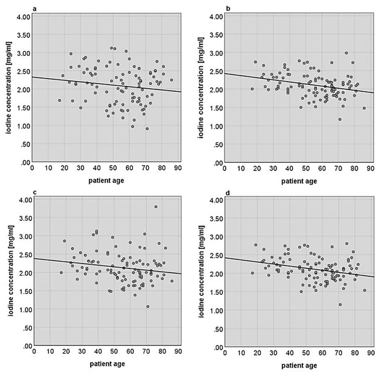
Dual energy computed tomography (DECT) allows the quantification of specific materials such as iodine contrast agent in human body tissue, potentially providing additional diagnostic data. Yet full diagnostic value can only be achieved if physiological normal values for iodine concentrations are known. We retrospectively evaluated abdominal DECT scans of 105 patients with healthy liver between March and August 2018 (age 17 to 86 years, 43 female and 62 male). The iodine concentrations within ROIs of the liver parenchyma as well as of the abdominal aorta and main portal vein were obtained. We evaluated the absolute iodine concentration and blood-normalized iodine concentrations relating the measured iodine concentration of the liver parenchyma to those of the supplying vessels. The influence of age and gender on the iodine uptake was assessed. The absolute iodine concentration was significantly different for the male and female cohort, but the difference was eliminated by the blood-normalized values. The average blood-normalized iodine concentrations were 2.107 mg/ml (+/- 0.322 mg/ml), 2.125 mg/ml (+/- 0.426 mg/ml) and 2.103 mg/ml (+/- 0.317 mg/ml) for the portal vein normalized, aorta normalized and mixed blood normalized iodine concentrations, respectively. A significant negative correlation between the patients' age and the iodine concentration was detected only for the blood-normalized values. A physiological range for iodine concentration in portal venous phase contrast enhanced DECT images can be defined for absolute and blood-normalized values. Deviations of blood-normalized iodine concentration values might be a robust biomarker for diagnostic evaluation. Patient age but not the gender influences the blood-normalized iodine concentrations in healthy liver parenchyma.


Conflict of interest statement

The authors have declared that no competing interests exist.

Figures

Fig 1
Fig 1. Dual energy CT iodine image, indicating the placement of the ROIs.
The 5 mm thick slice of a DECT iodine image in portal venous phase image at the level of the coeliac axis shows five ROIs, all with an approximate area of 1.5 cm2. Two ROIs (green) are placed in the right hepatic lobe in segments 4b and 7, one ROI (yellow) in the left hepatic lobe in segment 3, one ROI (blue) in the portal vein, and one in the aorta (red).
Fig 2
Fig 2. Quantitative iodine concentrations between genders.
(a) The absolute iodine concentrations IL_abs(x) show a significant difference (p < 0.001) between genders. The portal vein normalized iodine concentration Ipv-n(x) (b), the aorta normalized iodine concentration Ia-n(x) (c) and the mixed blood normalized iodine concentration Imixed-n(x) (d) do not show any significant differences between male and female patients.
Fig 3
Fig 3. Scatterplot diagrams relating the measured and calculated iodine concentrations to the patients’ age.
The measured absolute iodine concentration IL_abs(x) (a) does not show a significant correlation with the patients’ age (R2 = 0.021, r = -0.144, p = 0.080), while the different calculated blood-normalized iodine concentrations (b)–(d) do show a clear and significant negative correlation with the patients’ age. The portal vein normalized iodine concentration Ipv-n(x) (b) shows a correlation of R2 = 0.087 (r = -0.295, p < 0.01). The aorta normalized iodine concentration Ia-n(x) (c) shows a correlation of R2 = 0.031 (r = -0.177, p < 0.05). The mixed blood normalized iodine concentration Imixed -n(x) (d) shows a correlation of R2 = 0.087 (r = -0.295, p < 0.01).
Fig 4
Fig 4. Comparison of iodine uptake of young and elderly patient.
Fig (a) and (b) show the HU and iodine concentration DECT images of a young patient aged 20 years. Fig (c) and (d) show the HU and iodine concentration DECT images of an elderly patient aged 79 years. Although the iodine concentration in the portal vein of the young patient in fig (b) is lower than the iodine concentration in the portal vein of the elderly patient in fig (d), the iodine concentration of the liver parenchyma is visibly higher in the liver parenchyma of the younger patient (b) than it is for the elderly patient (d). The different uptake of contrast agent is clearly visible in the iodine images (b) and (d) while it is not that impressive in the HU images (a) and (c).

References

    1. Johnson TR. Dual-energy CT: general principles. AJR 2012; 199:S3–S8. doi: 10.2214/AJR.12.9116 - DOI - PubMed
    1. Coursey CA, Nelson RC, Boll DT, Paulson EK, Ho LM, Neville AM, et al.. Dual-Energy Multidetector CT: How Does It Work, What Can It Tell Us, and When Can We Use It in Abdominopelvic Imaging? Radiographics 2020; 30:1037–1055. 10.1148/rg.304095175 - DOI - PubMed
    1. Johnson TR, Kraus B, Sedlmair M, Grasruck M, Bruder H, Morhard D, et al.. Material differentiation by dual energy CT: initial experience, Eur Radiol 2007; 17:1510–1517. doi: 10.1007/s00330-006-0517-6 - DOI - PubMed
    1. Graser A, Johnson TR, Chandarana H, Macari M. Dual Energy CT: preliminary observations and potential clinical applications in the abdomen. Eur Radiol 2009; 19:13–23. doi: 10.1007/s00330-008-1122-7 - DOI - PubMed
    1. Johnson TR, Fink C, Schönberg SO, Reiser MF. Dual Energy CT in Clinical Practice, 1st ed. Springer Berlin Heidelberg New York, 2011.

Publication types