. 2022 Aug:82:104158.
doi: 10.1016/j.ebiom.2022.104158. Epub 2022 Jul 11.

Limited neutralisation of the SARS-CoV-2 Omicron subvariants BA.1 and BA.2 by convalescent and vaccine serum and monoclonal antibodies

Affiliations

Limited neutralisation of the SARS-CoV-2 Omicron subvariants BA.1 and BA.2 by convalescent and vaccine serum and monoclonal antibodies

Alexander Wilhelm et al. EBioMedicine. 2022 Aug.

Abstract

Background: In recent months, Omicron variants of SARS-CoV-2 have become dominant in many regions of the world, and case numbers with Omicron subvariants BA.1 and BA.2 continue to increase. Due to numerous mutations in the spike protein, the efficacy of currently available vaccines, which are based on Wuhan-Hu 1 isolate of SARS-CoV-2, is reduced, leading to breakthrough infections. Efficacy of monoclonal antibody therapy is also likely impaired.

Methods: In our in vitro study using A549-AT cells constitutively expressing ACE2 and TMPRSS2, we determined and compared the neutralizing capacity of vaccine-elicited sera, convalescent sera and monoclonal antibodies against authentic SARS-CoV-2 Omicron BA.1 and BA.2 compared with Delta.

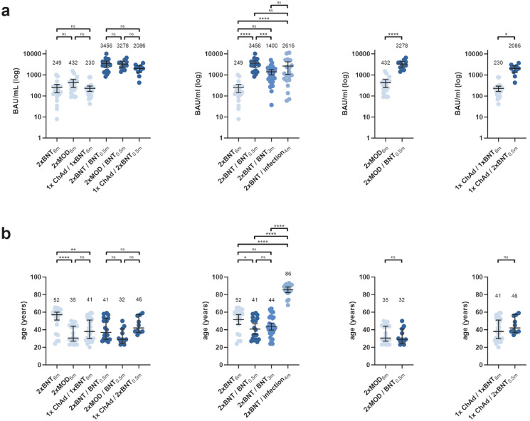
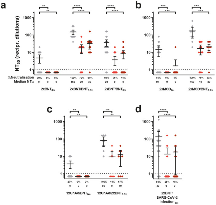
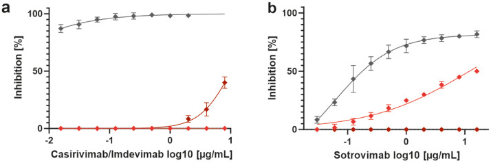
Findings: Almost no neutralisation of Omicron BA.1 and BA.2 was observed using sera from individuals vaccinated with two doses 6 months earlier, regardless of the type of vaccine taken. Shortly after the booster dose, most sera from triple BNT162b2-vaccinated individuals were able to neutralise both Omicron variants. In line with waning antibody levels three months after the booster, only weak residual neutralisation was observed for BA.1 (26%, n = 34, 0 median NT50) and BA.2 (44%, n = 34, 0 median NT50). In addition, BA.1 but not BA.2 was resistant to the neutralising monoclonal antibodies casirivimab/imdevimab, while BA.2 exhibited almost a complete evasion from the neutralisation induced by sotrovimab.

Interpretation: Both SARS-CoV-2 Omicron subvariants BA.1 and BA.2 escape antibody-mediated neutralisation elicited by vaccination, previous infection with SARS-CoV-2, and monoclonal antibodies. Waning immunity renders the majority of tested sera obtained three months after booster vaccination negative in BA.1 and BA.2 neutralisation. Omicron subvariant specific resistance to the monoclonal antibodies casirivimab/imdevimab and sotrovimab emphasizes the importance of genotype-surveillance and guided application.

Funding: This study was supported in part by the Goethe-Corona-Fund of the Goethe University Frankfurt (M.W.) and the Federal Ministry of Education and Research (COVIDready; grant 02WRS1621C (M.W.).

Keywords: BA.1; BA.2; Omicron; SARS-CoV-2; Sotrovimab; Waning Immunity.


Conflict of interest statement

Declaration of interests S.H. has received research support from Roche Diagnostics and a speaker's fee from Sanofi Genzyme. T.W. has received speaker and consultancy fees from Gilead Sciences, Merck Sharp & Dohme, and Janssen Pharmaceuticals. N.K. received speaker fees from Abbott. S.C. was a member of a clinical advisory board for Biontech. All other authors declare no conflicts of interest, financial or otherwise.

Figures

Figure 1:
Figure 1
Genotypic and phenotypic features of SARS-CoV-2 variants used in this study. a) Schematic drawing of SARS-CoV-2 BA.1 and BA.2 genomes compared to isolate Wuhan-Hu-1 (NC_045512) indicating spike positions common and unique within each of the indicated subvariant genome. The numbers denote nucleotide positions based on the reference strain NC_045512. ORFs based on reference sequence NC_045512 are shown as grey boxes. The receptor-binding domain (RBD), receptor-binding motif (RBM) as well as the N-terminal domain (NTD) are highlighted by green boxes. Heptad repeat domains 1 (HR1) and 2 (HR2), and the transmembrane region (TM) are indicated by orange boxes. Nucleotide substitutions compared to the reference sequence are indicated in the lower section. b-c) Representative cytopathic effect (CPE) formation and D) growth kinetics of SARS-CoV-2 variants in A549-AT cells (at least n = 12 biological replicates). CPE formation was analysed at b) peak syncytia formation and c) at the onset of cell lysis. Hours post infection (hpi). Scale bar in low-magnification images, 500 µm. Scale bar in high-magnification images, 180 µm. (For interpretation of the references to color in this figure legend, the reader is referred to the web version of this article.)
Figure 2
Figure 2
IgG antibody responses after SARS-CoV-2 booster vaccination and age distribution of the groups used in this study. a) SARS-CoV-2 IgG antibody concentrations were determined and indicated as binding antibody units per millilitre (BAU/mL). b) Age distribution of the serum donors grouped by vaccination scheme and sampling date. Bars indicate mean values with SD. BNT=BNT162b2; MOD=mRNA-1273; ChAd= ChAdOx1. Statistical significance was calculated by one-way ANOVA with Tukey's multiple comparison test. Asterisks indicate p-values as * (p < 0.05), ** (p < 0.01), *** (p < 0.001) and **** (p < 0.0001).
Figure 3
Figure 3
Antibody-mediated neutralisation of authentic SARS-CoV-2 Delta and Omicron subvariants BA.1 and BA.2. Values represent reciprocal dilutions micro-neutralisation titres resulting in 50% virus neutralisation (NT50) of SARS-CoV-2 variants Delta (grey) and Omicron BA.1 (red), BA.2 (dark red). a) Neutralisation assays were performed using serum samples obtained from vaccinated individuals receiving the indicated vaccine schemes (sampling time after last vaccination/booster indicated in subscript): double BNT162b2-vaccinated (2xBNT6m), and sera from triple BNT162b2-vaccinated individuals (2xBNT/BNT0.5m and 2xBNT/BNT3m) b) Neutralisation assays with sera from double mRNA-1273 vaccinated (2xMOD6m) and additionally BNT162b2-boosted (2xMOD/BNT0.5m) individuals. c) Neutralisation titres for sera from heterologous ChAdOx1 and BNT162b2-vaccinated (1xChAd/1xBNT6m) and BNT162b2-boosted (1xChAd/2xBNT0.5m) donors. d) Neutralisation assays performed with sera from double BNT162b2-vaccinated and SARS-CoV-2 infected individuals (2xBNT/SARS-CoV-2 infection4m). Information regarding the sera donors (sex, age, antibody titres test and time after vaccination) are summarised in Table 1 and Supplementary Figure 2. Median titres and relative portion (%) of neutralising sera against each variant are indicated below each panel. Mean values of two technical replicates per sample are depicted with 95% confidence intervals and SD. Statistical significance was calculated by two-tailed, Wilcoxon Rank Sum Test. Asterisks indicate p-values as * (p < 0.05), ** (p < 0.01), *** (p < 0.001) and **** (p < 0.0001). (For interpretation of the references to color in this figure legend, the reader is referred to the web version of this article.)
Figure 4
Figure 4
Waning antibody responses elicited by SARS-CoV-2 vaccination over time. a) Neutralising antibody responses were compared in BNT162b2-vaccinated individuals (2xBNT6m,n = 23; 2xBNT/BNT0.5m, n = 18; and 2xBNT/BNT3m, n = 34). Values represent reciprocal dilutions micro-neutralisation titres resulting in 50% virus neutralisation (NT50) of SARS-CoV-2 variants Delta (grey), Omicron BA.1 (red), and BA.2 (dark red). Median titres and relative portion (%) of neutralising sera against each variant are indicated above or below each panel, respectively. Mean values of two technical replicates per sample are depicted. b) Decrease in variant-specific neutralisation titres from panel A over time. Statistical significance was calculated by multivariate analysis including demographic covariates (Supplementary Table 4). Asterisks indicate p-values as * (p < 0.05), ** (p < 0.01) and *** (p < 0.001). (For interpretation of the references to color in this figure legend, the reader is referred to the web version of this article.)
Figure 5
Figure 5
Neutralisation of SARS-CoV-2 subvariants by mAbs. Comparison of EC50 values by monoclonal antibodies a) imdevimab/casirivimab (applied in a 1:1 ratio) and b) sotrovimab against parental SARS-CoV-2 strain B/FFM5 (grey), BA.1 (red), and BA.2 (dark red). Experiments were performed in three biological replicates using A549-AT cells. Readout was performed two days after infection. Bars indicate mean values with SD. The dotted line indicates the upper quantification limit of the assay. (For interpretation of the references to color in this figure legend, the reader is referred to the web version of this article.)

References

    1. Zhou P, Yang XL, Wang XG, et al. A pneumonia outbreak associated with a new coronavirus of probable bat origin. Nature. 2020;579(7798):270–273. - PMC - PubMed
    1. Zhu N, Zhang D, Wang W, et al. A novel Coronavirus from patients with pneumonia in China, 2019. N Engl J Med. 2020;382(8):727–733. - PMC - PubMed
    1. WHO . Emergency Situation Updates. World Health Organisation (WHO); 2022. COVID-19 Weekly Epidemiological Update on COVID-19.
    1. Hoffmann M, Kleine-Weber H, Schroeder S, et al. SARS-CoV-2 cell entry depends on ACE2 and TMPRSS2 and is blocked by a clinically proven protease inhibitor. Cell. 2020;181(2):271–280.e8. - PMC - PubMed
    1. Harvey WT, Carabelli AM, Jackson B, et al. SARS-CoV-2 variants, spike mutations and immune escape. Nat Rev Microbiol. 2021;19(7):409–424. - PMC - PubMed

Supplementary concepts