

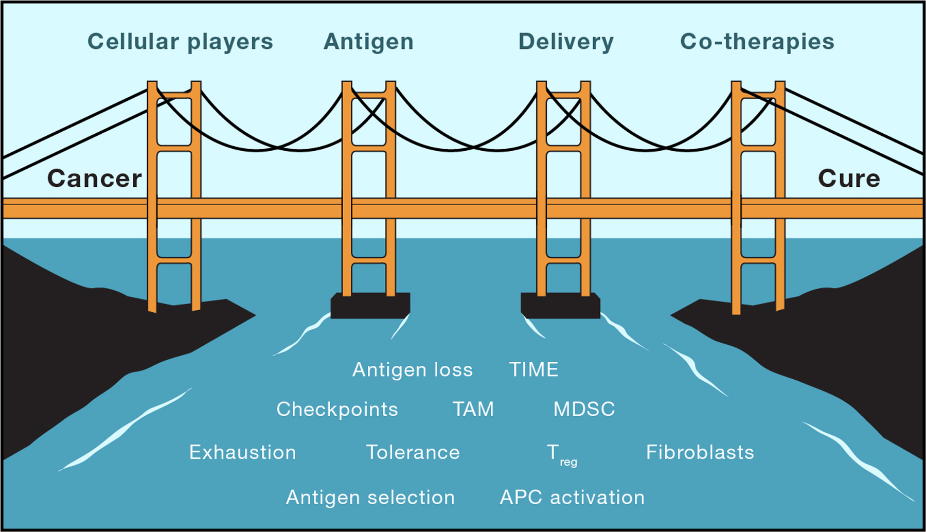
Cancer vaccines: Building a bridge over troubled waters
- PMID: 35835100
- PMCID: PMC9555301
- DOI: 10.1016/j.cell.2022.06.035
Cancer vaccines: Building a bridge over troubled waters
Abstract
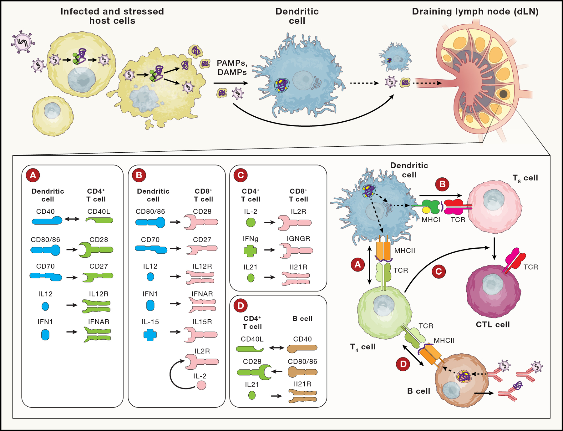
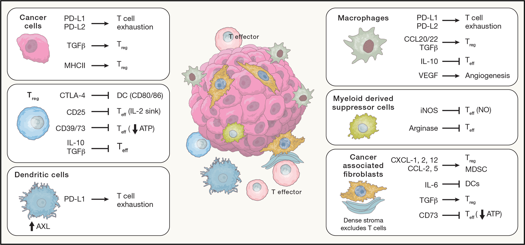
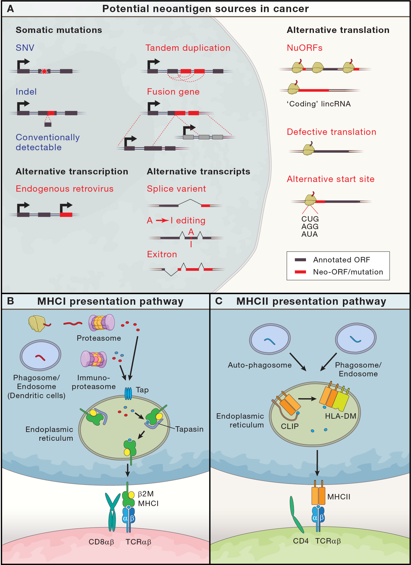
Cancer vaccines aim to direct the immune system to eradicate cancer cells. Here we review the essential immunologic concepts underpinning natural immunity and highlight the multiple unique challenges faced by vaccines targeting cancer. Recent technological advances in mass spectrometry, neoantigen prediction, genetically and pharmacologically engineered mouse models, and single-cell omics have revealed new biology, which can help to bridge this divide. We particularly focus on translationally relevant aspects, such as antigen selection and delivery and the monitoring of human post-vaccination responses, and encourage more aggressive exploration of novel approaches.
Copyright © 2022 Elsevier Inc. All rights reserved.
Conflict of interest statement
Declaration of interests E.F.F. is an equity holder in and consultant for BioNTech, an equity holder and scientific advisory board member of BioEntre, and a founder and equity holder of Dionis Therapeutics. C.J.W. is an equity holder in BioNTech. An immediate family member of C.J.W. is an advisor and equity holder for Related Sciences and receives research funding from Bristol-Myers Squibb. Patent applications have been filed that relate to the reviewed material, as follows: “compositions and methods for personalized neoplasia vaccines” (E.F.F. and C.J.W.), “methods for identifying tumor-specific neoantigens” (C.J.W.), “formulations for neoplasia vaccines” (E.F.F.), “combination therapy for neoantigen vaccine” (C.J.W. and E.F.F.), and “multi-domain protein vaccine” (E.F.F.).
Figures
References
-
- Abdel-Hakeem MS, Manne S, Beltra JC, Stelekati E, Chen Z, Nzingha K, Ali MA, Johnson JL, Giles JR, Mathew D, Greenplate AR, Vahedi G, and Wherry EJ (2021). Epigenetic scarring of exhausted T cells hinders memory differentiation upon eliminating chronic antigenic stimulation. Nat Immunol 22, 1008–1019. 10.1038/s41590-021-00975-5. - DOI - PMC - PubMed
-
- Abelin JG, Harjanto D, Malloy M, Suri P, Colson T, Goulding SP, Creech AL, Serrano LR, Nasir G, Nasrullah Y, McGann CD, Velez D, Ting YS, Poran A, Rothenberg DA, Chhangawala S, Rubinsteyn A, Hammerbacher J, Gaynor RB, Fritsch EF, Greshock J, Oslund RC, Barthelme D, Addona TA, Arieta CM, and Rooney MS (2019). Defining HLA-II Ligand Processing and Binding Rules with Mass Spectrometry Enhances Cancer Epitope Prediction. Immunity 51, 766–779 e717. 10.1016/j.immuni.2019.08.012. - DOI - PubMed
-
- Abusarah J, Khodayarian F, El-Hachem N, Salame N, Olivier M, Balood M, Roversi K, Talbot S, Bikorimana JP, Chen J, Jolicoeur M, Trudeau LE, Kamyabiazar S, Annabi B, Robert F, Pelletier J, El-Kadiry AE, Shammaa R, and Rafei M (2021). Engineering immunoproteasome-expressing mesenchymal stromal cells: A potent cellular vaccine for lymphoma and melanoma in mice. Cell Rep Med 2, 100455. 10.1016/j.xcrm.2021.100455. - DOI - PMC - PubMed
-
- Alspach E, Lussier DM, Miceli AP, Kizhvatov I, DuPage M, Luoma AM, Meng W, Lichti CF, Esaulova E, Vomund AN, Runci D, Ward JP, Gubin MM, Medrano RFV, Arthur CD, White JM, Sheehan KCF, Chen A, Wucherpfennig KW, Jacks T, Unanue ER, Artyomov MN, and Schreiber RD (2019). MHC-II neoantigens shape tumour immunity and response to immunotherapy. Nature 574, 696–701. 10.1038/s41586-019-1671-8. - DOI - PMC - PubMed
Publication types
MeSH terms
Substances
Grants and funding
LinkOut - more resources
Full Text Sources
Medical
