

Spontaneous NLRP3 inflammasome-driven IL-1-β secretion is induced in severe COVID-19 patients and responds to anakinra treatment
- PMID: 35835255
- PMCID: PMC9272569
- DOI: 10.1016/j.jaci.2022.05.029
Spontaneous NLRP3 inflammasome-driven IL-1-β secretion is induced in severe COVID-19 patients and responds to anakinra treatment
Abstract
Background: Severe acute respiratory syndrome coronavirus 2 (SARS-CoV-2) infection may result in a severe pneumonia associated with elevation of blood inflammatory parameters, reminiscent of cytokine storm syndrome. Steroidal anti-inflammatory therapies have shown efficacy in reducing mortality in critically ill patients; however, the mechanisms by which SARS-CoV-2 triggers such an extensive inflammation remain unexplained.
Objectives: To dissect the mechanisms underlying SARS-CoV-2-associated inflammation in patients with severe coronavirus disease 2019 (COVID-19), we studied the role of IL-1β, a pivotal cytokine driving inflammatory phenotypes, whose maturation and secretion are regulated by inflammasomes.
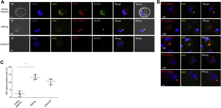
Methods: We analyzed nod-like receptor protein 3 pathway activation by means of confocal microscopy, plasma cytokine measurement, cytokine secretion following in vitro stimulation of blood circulating monocytes, and whole-blood RNA sequencing. The role of open reading frame 3a SARS-CoV-2 protein was assessed by confocal microscopy analysis following nucleofection of a monocytic cell line.
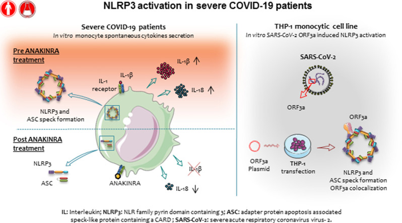
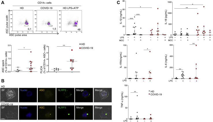
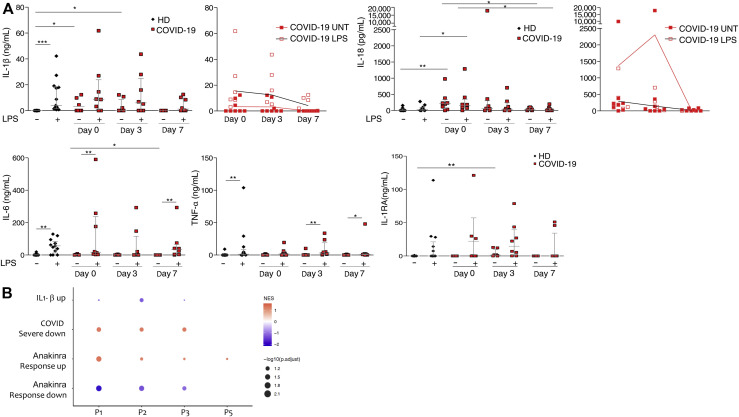
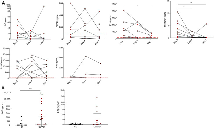
Results: We found that circulating monocytes from patients with COVID-19 display ASC (adaptor molecule apoptotic speck like protein-containing a CARD) specks that colocalize with nod-like receptor protein 3 inflammasome and spontaneously secrete IL-1β in vitro. This spontaneous activation reverts following patient's treatment with the IL-1 receptor antagonist anakinra. Transfection of a monocytic cell line with cDNA coding for the ORF3a SARS-CoV-2 protein resulted in ASC speck formation.
Conclusions: These results provide further evidence that IL-1β targeting could represent an effective strategy in this disease and suggest a mechanistic explanation for the strong inflammatory manifestations associated with COVID-19.
Keywords: IL-1β; NLRP3 inflammasome; SARS-CoV-2; inflammation.
Copyright © 2022 American Academy of Allergy, Asthma & Immunology. Published by Elsevier Inc. All rights reserved.
Figures
Similar articles
-
Targeting the NLRP3 Inflammasome in Severe COVID-19.Front Immunol. 2020 Jun 23;11:1518. doi: 10.3389/fimmu.2020.01518. eCollection 2020. Front Immunol. 2020. PMID: 32655582 Free PMC article. Review.
-
Severe acute respiratory syndrome coronavirus ORF3a protein activates the NLRP3 inflammasome by promoting TRAF3-dependent ubiquitination of ASC.FASEB J. 2019 Aug;33(8):8865-8877. doi: 10.1096/fj.201802418R. Epub 2019 Apr 29. FASEB J. 2019. PMID: 31034780 Free PMC article.
-
Interleukin-1 and the NLRP3 inflammasome in COVID-19: Pathogenetic and therapeutic implications.EBioMedicine. 2022 Nov;85:104299. doi: 10.1016/j.ebiom.2022.104299. Epub 2022 Oct 6. EBioMedicine. 2022. PMID: 36209522 Free PMC article. Review.
-
The NLRP3 inflammasome and COVID-19: Activation, pathogenesis and therapeutic strategies.Cytokine Growth Factor Rev. 2021 Oct;61:2-15. doi: 10.1016/j.cytogfr.2021.06.002. Epub 2021 Jun 18. Cytokine Growth Factor Rev. 2021. PMID: 34183243 Free PMC article. Review.
-
SARS-CoV-2 viroporin encoded by ORF3a triggers the NLRP3 inflammatory pathway.Virology. 2022 Mar;568:13-22. doi: 10.1016/j.virol.2022.01.003. Epub 2022 Jan 17. Virology. 2022. PMID: 35066302 Free PMC article.
Cited by
-
Sarcoidosis and COVID-19: At the Cross-Road between Immunopathology and Clinical Manifestation.Biomedicines. 2022 Oct 9;10(10):2525. doi: 10.3390/biomedicines10102525. Biomedicines. 2022. PMID: 36289785 Free PMC article. Review.
-
COVID-19 and trained immunity: the inflammatory burden of long covid.Front Immunol. 2023 Nov 28;14:1294959. doi: 10.3389/fimmu.2023.1294959. eCollection 2023. Front Immunol. 2023. PMID: 38090572 Free PMC article.
-
NLRP-3 Inflammasome: A Key Target, but Mostly Overlooked following SARS-CoV-2 Infection.Vaccines (Basel). 2022 Aug 12;10(8):1307. doi: 10.3390/vaccines10081307. Vaccines (Basel). 2022. PMID: 36016195 Free PMC article.
-
Inflammasomes: a rising star on the horizon of COVID-19 pathophysiology.Front Immunol. 2023 May 12;14:1185233. doi: 10.3389/fimmu.2023.1185233. eCollection 2023. Front Immunol. 2023. PMID: 37251383 Free PMC article. Review.
-
Autosomal Dominant Polycystic Kidney Disease-Related Multifocal Renal Cell Carcinoma: A Narrative Iconographic Review.Int J Mol Sci. 2025 Apr 23;26(9):3965. doi: 10.3390/ijms26093965. Int J Mol Sci. 2025. PMID: 40362206 Free PMC article. Review.
References
Publication types
MeSH terms
Substances
LinkOut - more resources
Full Text Sources
Molecular Biology Databases
Miscellaneous
