. 2022 Aug 1;72(4):273-279.
doi: 10.30802/AALAS-CM-21-000107. Epub 2022 Jul 14.

Use of Fluconazole-impregnated Beads to Treat Osteomyelitis Caused by Coccidioides in a Pigtailed Macaque (Macaca nemestrina)

Affiliations

Use of Fluconazole-impregnated Beads to Treat Osteomyelitis Caused by Coccidioides in a Pigtailed Macaque (Macaca nemestrina)

Charlotte E Hotchkiss et al. Comp Med. .

Abstract

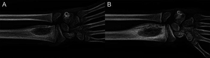
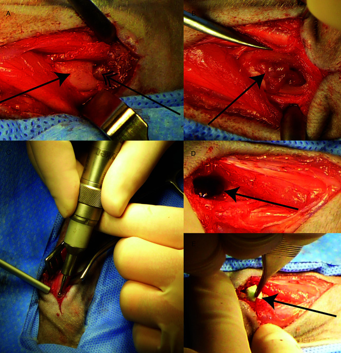
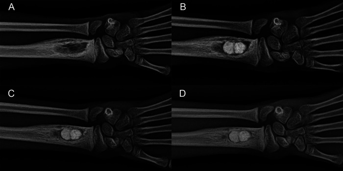
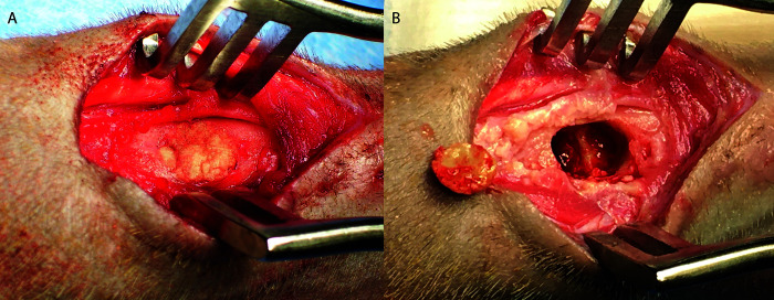
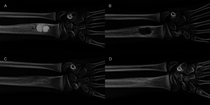
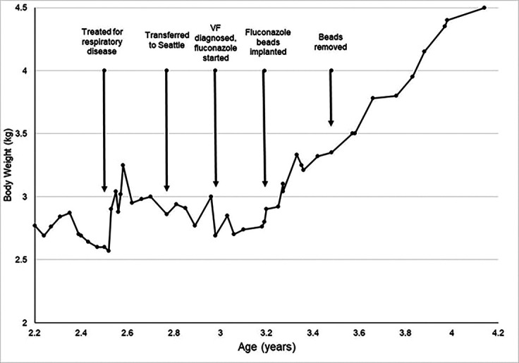
A 3-y-old male pigtailed macaque (Macaca nemestrina) presented for swelling of the left distal forearm and decreased use of the arm. The monkey had been raised at an indoor-outdoor facility in Arizona and transferred to an indoor facility in Washington 2 mo prior to presentation. A preliminary diagnosis of fungal osteomyelitis of the radius was made based on radiographs and Coccidioides titers. In addition to systemic antifungal treatment, surgery was performed to debride the bony lesion and implant polymethylmethacrylate beads impregnanted with the anti-fungal fluconazole. Histologic examination of the debrided material confirmed the diagnosis of fungal osteomyelitis. The surgical procedure resulted in clinical improvement, as evidenced by weight gain and decreased Coccidioides titers. The beads were removed in a second surgery, and the bony lesion completely resolved. With continued systemic fluconazole treatment, the monkey remained healthy with no further evidence of osteomyelitis. Coccidioides is an emerging pathogen that causes significant morbidity and mortality in both humans and animals. Bone infections can be resistant to systemic treatment, and the implantation of fluconazoleimpregnated beads may offer a successful treatment strategy for fungal osteomyelitis.


Figures

Figure 1.
Figure 1.
Fungal osteomyelitis in the distal radius of a M. nemestrina. A) Initial presentation. A single osteolytic lesion is seen within the left radius, with cortical thinning and periosteal reaction on the lateral surface. B) After 4 wk of systemic fluconazole treatment, there was increased periosteal proliferation with loss of cortical definition on the lateral surface, irregular density within the lesion, and decreased definition at the margins of the lesion.
Figure 2.
Figure 2.
Bead implantation surgery. A) Incision over dorsal surface of distal radius showing both bony and soft tissue swelling. Solid arrow = radius bone, double arrow = soft tissue in bony defect. B) Pyogranulomatous tissue with reactive fibroplasia (arrow) partially removed from the medullary cavity of the distal radius, C) Expansion of the cortical bone defect using a surgical drill, D) Medullary cavity (arrow) after debridement, E) Fluconazole-impregnated PMMA beads (arrow) within the medullary cavity.
Figure 3.
Figure 3.
Radiographs associated with bead insertion. A) Preoperative, approximately 10 wk after initial presentation. After additional systemic fluconazole treatment while preparing for surgery, the periosteal reaction appears to be resolving. However, the osteolytic lesion in the radius is unchanged in size. B) Immediately after surgery. Placement of 2 fluconazole-impregnated beads within the radius was confirmed. An area of radiolucency is present proximal to the beads. C) 4 wk after implantation. Radiodensity has increased around the beads. D) 8 wk after implantation. Radiodensity has further increased around the beads and has the appearance of trabecular bone.
Figure 4.
Figure 4.
Bead removal surgery. A) Beads visible in situ within radius, B) Partial bead that had been removed shown next to cavity after bead removal. Note ridge in center of cavity where bone had remodeled around beads.
Figure 5.
Figure 5.
Radiographs associated with bead removal. A) Preoperative on the day of bead removal, 15 wk after the initial surgery. Beads appear unchanged and are surrounded by trabecular bone. B) 4 wk after bead removal. The void where the beads were persists, but the surrounding bone has normal trabecular architecture. C) 4 mo after bead removal. The defect where the beads had been has closed over. Some irregularity of the cortical/trabecular margins persists. D) 8 mo after bead removal. The bone shows normal trabecular architecture and smooth cortical contours.
Figure 6.
Figure 6.
Body weight changes over time in relation to clinical events. VF = Valley Fever. A juvenile M. nemestrina is expected to steadily gain weight between 2 and 4 y of age. Weight gain stopped during clinical respiratory illness but rebounded after symptomatic treatment. Weight gain stopped again around the time of transfer to Seattle, and the diagnosis of VF was made after weight loss. Systemic treatment with fluconazole stabilized weight but weight gain still did not occur. After surgery to debride the fungal bony lesion and implant fluconazole-impregnated beads, normal weight gain resumed. Weight gain continued uninterrupted after surgical removal of the beads.

Similar articles

Cited by

References

    1. Abosala A, Ali M. 2020. The Use of Calcium Sulphate beads in Periprosthetic Joint Infection, a systematic review. J Bone Jt Infect 5:43–49. 10.7150/jbji.41743. - DOI - PMC - PubMed
    1. Anagnostakos K, Kelm J, Schmitt E, Jung J. 2012. Fungal Periprosthetic Hip and Knee Joint Infections: Clinical Experience With a 2-Stage Treatment Protocol. J Arthroplasty 27:293–298. 10.1016/j.arth.2011.04.044. - DOI - PubMed
    1. Arbona N, Butkiewicz CD, Keyes M, Shubitz LF. 2019. Clinical features of cats diagnosed with coccidioidomycosis in Arizona, 2004-2018. J Feline Med Surg 22:129–137. 10.1177/1098612x19829910. - DOI - PMC - PubMed
    1. Beaman L, Holmberg C, Henrickson R, Osburn B. 1980. The incidence of coccidioidomycosis among nonhuman primates housed outdoors at the California Primate Research Center. J Med Primatol 9:254–261. 10.1159/000460147. - DOI - PubMed
    1. Blundell GP, Castleberry MW, Lowe EP, Converse JL. 1961. The pathology of Coccidioides immitis in the Macaca mulatta. Am J Pathol 39:613–630. - PMC - PubMed

Publication types