

Multimodal Applications of 3D-Navigation in Single-Level Minimally Invasive Transforaminal Lumbar Interbody Fusion: Impacts on Precision, Accuracy, Complications, and Radiation Exposure
- PMID: 35835566
- PMCID: PMC9421208
- DOI: 10.14444/8294
Multimodal Applications of 3D-Navigation in Single-Level Minimally Invasive Transforaminal Lumbar Interbody Fusion: Impacts on Precision, Accuracy, Complications, and Radiation Exposure
Abstract
Background: Three-dimensional (3D)-navigation in minimally invasive transforaminal lumbar interbody fusion (MI-TLIF) is an evolving procedure. It is used not only for its accuracy of pedicle screw fixation but also for other major steps in transforaminal lumbar interbody fusion. Multimodal outcomes of this procedure are very limited in the literature. The purpose of this study was to examine the application of 3D-navigation in minimally invasive transforaminal lumbar interbody fusion (MI-TLIF).
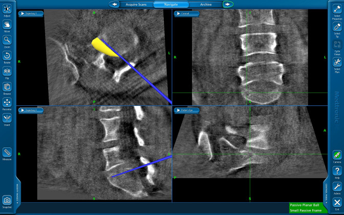
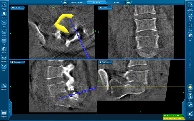
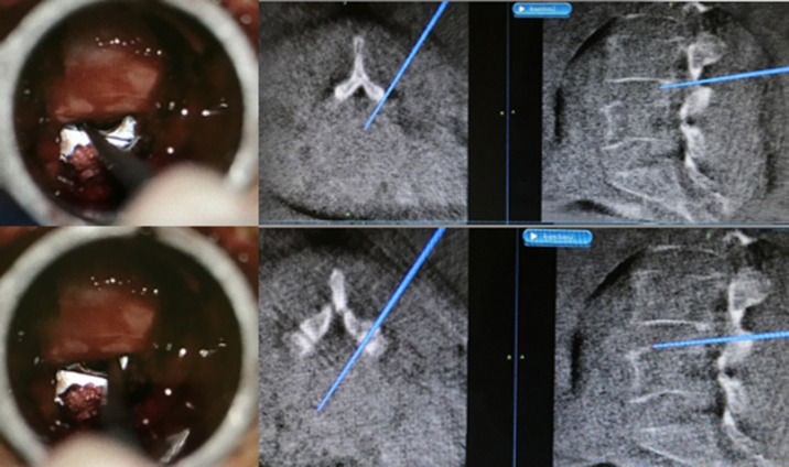
Methods: Patients who underwent single-level MI-TLIF using 3D-navigation between January 2017 and July 2019 were evaluated for navigation setting time, radiation exposure, volume of nucleus pulposus excised, cage placement, accuracy of pedicle screw placement, and cranial facet-joint violation.
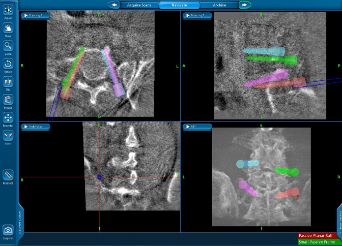
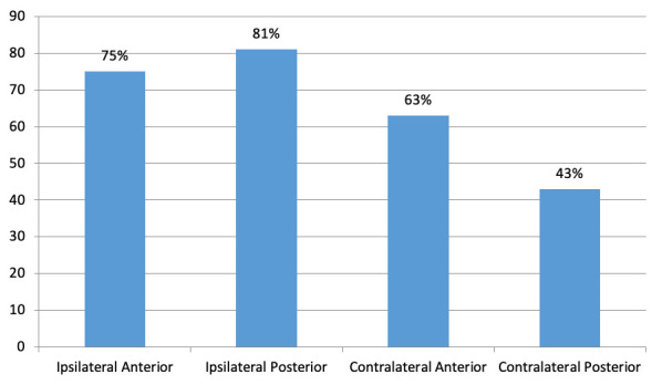
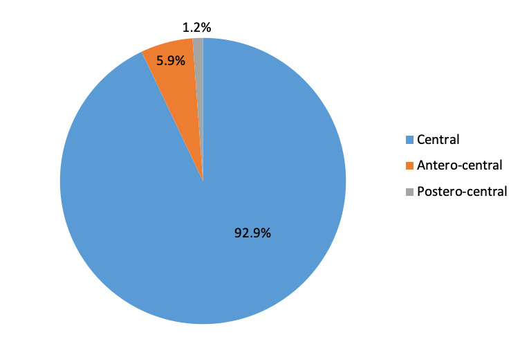
Results: One hundred and two patients with a mean age of 60.2 years met the inclusion criteria. The mean presetting time of navigation was 46.65 ± 9.45 minutes. Radiation exposure, fluoroscopy use, and fluoroscopy time were 15.54 ± 0.65 mGy, 4.43 ± 0.87 Gy.cm², and 97.6 ± 11.67 seconds, respectively. The mean amount of nucleus pulposus excised from all quadrants was quantified. The cage was centrally placed in 87 patients, with 95.4% showing a Grade 0 pedicle breach and 94.6% showing Grade 0 cranial facet-joint violation.
Conclusion: Registration and setting up 3D-navigation takes additional time. The amount of exposure to the patient is much less compared to routine computed tomography, and, importantly, the operating team is protected from radiation. Navigated MI-TLIF has high rates of accuracy with regard to placement of percutaneous pedicle screws and cages with the added advantage of protection of the cranial facet-joint.
Keywords: 3D-navigation; lumbar spine; minimally invasive spine surgery; spinal navigation; transforaminal lumbar interbody fusion.
This manuscript is generously published free of charge by ISASS, the International Society for the Advancement of Spine Surgery. Copyright © 2022 ISASS. To see more or order reprints or permissions, see http://ijssurgery.com.
Conflict of interest statement
Declaration of Conflicting Interests: The authors report no conflicts of interest in this work.
Figures

References
LinkOut - more resources
Full Text Sources
Miscellaneous