. 2023 Jan;64(1):96-101.
doi: 10.2967/jnumed.122.264112. Epub 2022 Jul 14.

Optimized Whole-Body PET MRI Sequence Workflow in Pediatric Hodgkin Lymphoma Patients

Affiliations

Optimized Whole-Body PET MRI Sequence Workflow in Pediatric Hodgkin Lymphoma Patients

Thomas W Georgi et al. J Nucl Med. 2023 Jan.

Abstract

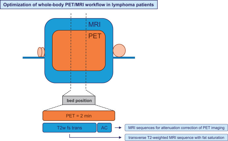
18F-FDG PET/MRI might be the diagnostic method of choice for Hodgkin lymphoma patients, as it combines significant metabolic information from PET with excellent soft-tissue contrast from MRI and avoids radiation exposure from CT. However, a major issue is longer examination times than for PET/CT, especially for younger children needing anesthesia. Thus, a targeted selection of suitable whole-body MRI sequences is important to optimize the PET/MRI workflow. Methods: The initial PET/MRI scans of 84 EuroNet-PHL-C2 study patients from 13 international PET centers were evaluated. In each available MRI sequence, 5 PET-positive lymph nodes were assessed. If extranodal involvement occurred, 2 splenic lesions, 2 skeletal lesions, and 2 lung lesions were also assessed. A detection rate was calculated dividing the number of visible, anatomically assignable, and measurable lesions in the respective MRI sequence by the total number of lesions. Results: Relaxation time-weighted (T2w) transverse sequences with fat saturation (fs) yielded the best result, with detection rates of 95% for nodal lesions, 62% for splenic lesions, 94% for skeletal lesions, and 83% for lung lesions, followed by T2w transverse sequences without fs (86%, 49%, 16%, and 59%, respectively) and longitudinal relaxation time-weighted contrast-enhanced transverse sequences with fs (74%, 35%, 57%, and 55%, respectively). Conclusion: T2w transverse sequences with fs yielded the highest detection rates and are well suited for accurate whole-body PET/MRI in lymphoma patients. There is no evidence to recommend the use of contrast agents.

Keywords: Hodgkin lymphoma; MRI sequences; PET/MRI; whole-body imaging.


Figures

None
Graphical abstract
FIGURE 1.
FIGURE 1.
Maximum-intensity projection of PET image of study patient with nodal, splenic, skeletal, and lung lesions. Scale bars represent SUV (unitless).
FIGURE 2.
FIGURE 2.
Corresponding transverse slice positions of PET and PET/MRI. (A and C) Transverse thoracic slice with PET-positive right-sided internal mammary lymph node (green arrow) and PET-positive right-sided hilar lymph node (red arrow). (B and D) Transverse abdominal slice with 2 PET-positive splenic lesions (green and red arrow).
FIGURE 3.
FIGURE 3.
Identical transverse slice positions of different MRI sequences (same slice position as in Figs. 2A and 2C). (A) T1w MRI sequence with ce. (B) T2w MRI sequence with fs. (C) T2w nonfs MRI sequence. (D) Dixon relative water fraction distribution. PET-positive right-sided internal mammary lymph node (green arrow) is visible in A, B, and C. PET-positive right-sided hilar lymph node (red arrow) is visible in A, B, and D.
FIGURE 4.
FIGURE 4.
Identical transverse slice positions of different MRI sequences (same slice position as in Figs. 2B and 2D). (A) T1w MRI sequence with ce. (B) T2w MRI sequence with fs. (C) T2w nonfs MRI sequence. (D) Dixon relative water fraction distribution. Dorsal splenic lesion (red arrow) is visible in A–D; ventral lesion, only in B (green arrow).

Similar articles

Cited by

References

    1. Verhagen MV, Menezes LJ, Neriman D, et al. . 18F-FDG PET/MRI for staging and interim response assessment in pediatric and adolescent Hodgkin lymphoma: a prospective study with 18F-FDG PET/CT as the reference standard. J Nucl Med. 2021;62:1524–1530. - PMC - PubMed
    1. Herrmann K, Queiroz M, Huellner MW, et al. . Diagnostic performance of FDG-PET/MRI and WB-DW-MRI in the evaluation of lymphoma: a prospective comparison to standard FDG-PET/CT. BMC Cancer. 2015;15:1002. - PMC - PubMed
    1. Spick C, Herrmann K, Czernin J. 18F-FDG PET/CT and PET/MRI perform equally well in cancer: evidence from studies on more than 2,300 patients. J Nucl Med. 2016;57:420–430. - PMC - PubMed
    1. Ferdová E, Ferda J, Baxa J. 18F-FDG-PET/MRI in lymphoma patients. Eur J Radiol. 2017;94:A52–A63. - PubMed
    1. Huang B, Law MW, Khong PL. Whole‐body PET/CT scanning: estimation of radiation dose and cancer risk. Radiology. 2009;251:166–174. - PubMed

Publication types

Substances