

The deciphering of the immune cells and marker signature in COVID-19 pathogenesis: An update
- PMID: 35835586
- PMCID: PMC9350195
- DOI: 10.1002/jmv.28000
The deciphering of the immune cells and marker signature in COVID-19 pathogenesis: An update
Abstract
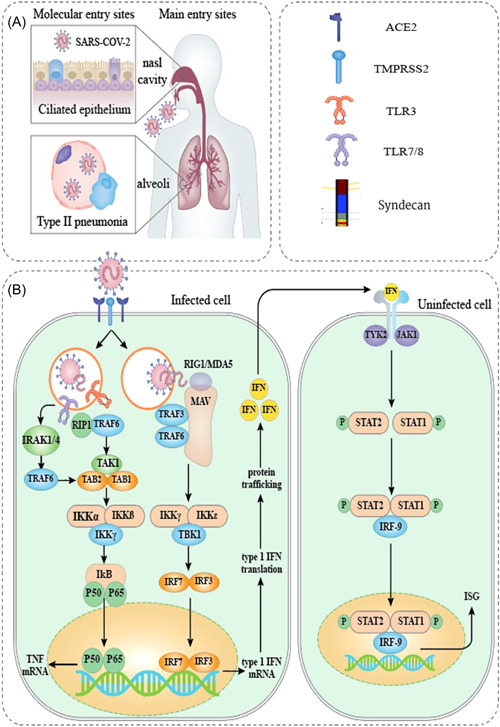
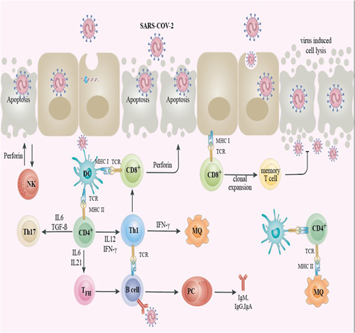
The precise interaction between the immune system and severe acute respiratory syndrome coronavirus 2 (SARS-CoV-2) is critical in deciphering the pathogenesis of coronavirus disease 2019 (COVID-19) and is also vital for developing novel therapeutic tools, including monoclonal antibodies, antivirals drugs, and vaccines. Viral infections need innate and adaptive immune reactions since the various immune components, such as neutrophils, macrophages, CD4+ T, CD8+ T, and B lymphocytes, play different roles in various infections. Consequently, the characterization of innate and adaptive immune reactions toward SARS-CoV-2 is crucial for defining the pathogenicity of COVID-19. In this study, we explain what is currently understood concerning the conventional immune reactions to SARS-CoV-2 infection to shed light on the protective and pathogenic role of immune response in this case. Also, in particular, we investigate the in-depth roles of other immune mediators, including neutrophil elastase, serum amyloid A, and syndecan, in the immunopathogenesis of COVID-19.
Keywords: COVID-19; adaptive immunity; innate immunity; neutrophil elastase; pathogenesis; syndecan.
© 2022 Wiley Periodicals LLC.
Conflict of interest statement
The authors declare no conflict of interest.
Figures
Similar articles
-
Immune response induced by novel coronavirus infection.Front Cell Infect Microbiol. 2022 Oct 25;12:988604. doi: 10.3389/fcimb.2022.988604. eCollection 2022. Front Cell Infect Microbiol. 2022. PMID: 36389144 Free PMC article. Review.
-
Influence of Aerosol Delivered BCG Vaccination on Immunological and Disease Parameters Following SARS-CoV-2 Challenge in Rhesus Macaques.Front Immunol. 2022 Feb 9;12:801799. doi: 10.3389/fimmu.2021.801799. eCollection 2021. Front Immunol. 2022. PMID: 35222355 Free PMC article.
-
Innate and adaptive immunity to SARS-CoV-2 and predisposing factors.Front Immunol. 2023 May 9;14:1159326. doi: 10.3389/fimmu.2023.1159326. eCollection 2023. Front Immunol. 2023. PMID: 37228604 Free PMC article. Review.
-
The laboratory tests and host immunity of COVID-19 patients with different severity of illness.JCI Insight. 2020 May 21;5(10):e137799. doi: 10.1172/jci.insight.137799. JCI Insight. 2020. PMID: 32324595 Free PMC article.
-
A comprehensive review about immune responses and exhaustion during coronavirus disease (COVID-19).Cell Commun Signal. 2022 Jun 2;20(1):79. doi: 10.1186/s12964-022-00856-w. Cell Commun Signal. 2022. PMID: 35655192 Free PMC article. Review.
Cited by
-
Drug repurposing of pyrazolotriazine derivatives as potential anti-SARS-CoV-2 agents: in vitro and in silico studies.BMC Chem. 2024 Jul 16;18(1):132. doi: 10.1186/s13065-024-01233-z. BMC Chem. 2024. PMID: 39014447 Free PMC article.
-
The central role of neutrophil extracellular traps (NETs) and by-products in COVID-19 related pulmonary thrombosis.Immun Inflamm Dis. 2023 Aug;11(8):e949. doi: 10.1002/iid3.949. Immun Inflamm Dis. 2023. PMID: 37647446 Free PMC article. Review.
-
Dynamics of Innate Immunity in SARS-CoV-2 Infections: Exploring the Impact of Natural Killer Cells, Inflammatory Responses, Viral Evasion Strategies, and Severity.Cells. 2025 May 22;14(11):763. doi: 10.3390/cells14110763. Cells. 2025. PMID: 40497938 Free PMC article. Review.
-
Serum IFN-γ and RNAemia temporal profiles as biomarkers of severe COVID-19 in solid organ transplant and immunocompetent patients.J Infect. 2023 May;86(5):529-533. doi: 10.1016/j.jinf.2023.01.019. Epub 2023 Jan 21. J Infect. 2023. PMID: 36690212 Free PMC article. No abstract available.
-
Regulatory T Cells (Tregs) and COVID-19: Unveiling the Mechanisms, and Therapeutic Potentialities with a Special Focus on Long COVID.Vaccines (Basel). 2023 Mar 19;11(3):699. doi: 10.3390/vaccines11030699. Vaccines (Basel). 2023. PMID: 36992283 Free PMC article. Review.
References
Publication types
MeSH terms
LinkOut - more resources
Full Text Sources
Medical
Research Materials
Miscellaneous
