

Bioconjugation Strategies for Tobacco Mild Green Mosaic Virus
- PMID: 35835718
- PMCID: PMC9624232
- DOI: 10.1002/cbic.202200323
Bioconjugation Strategies for Tobacco Mild Green Mosaic Virus
Abstract
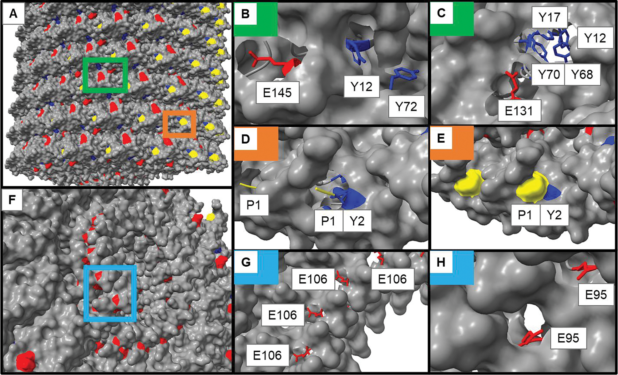
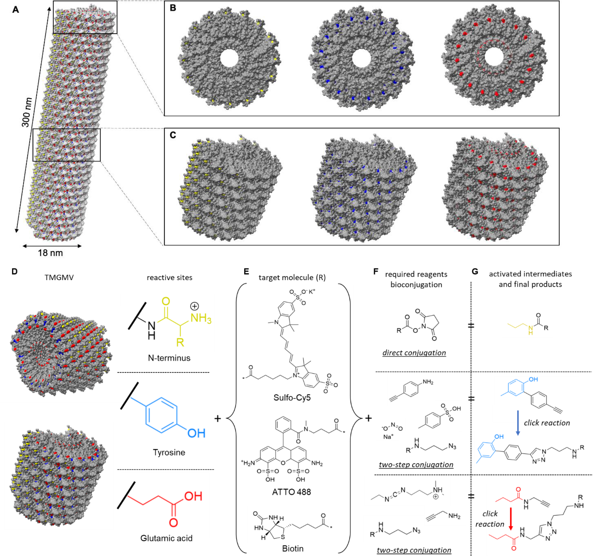
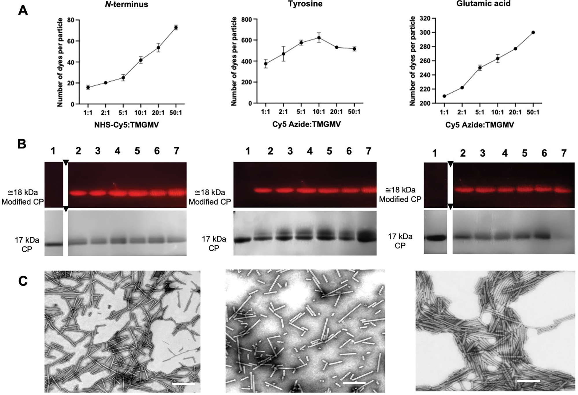
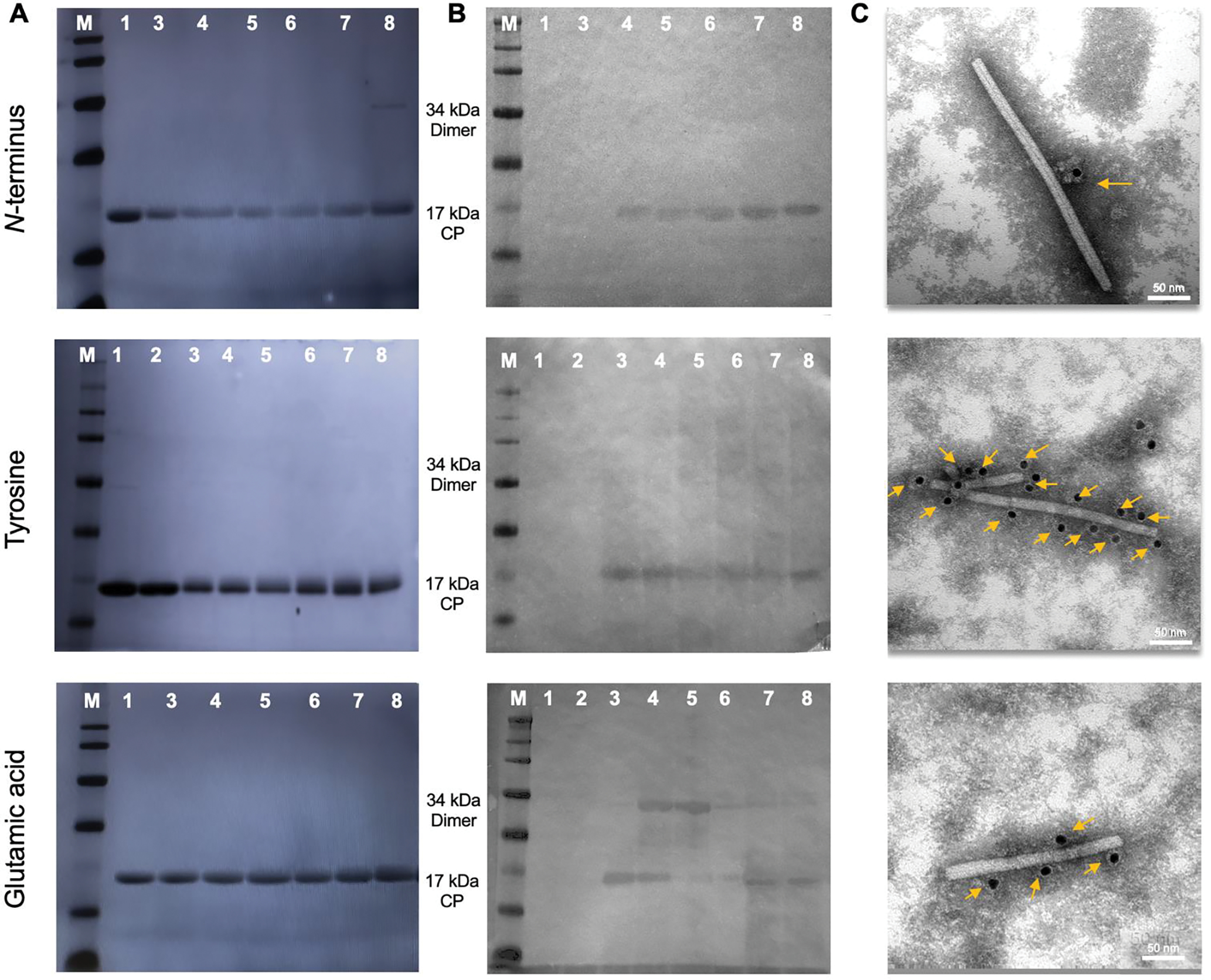
Tobacco mild green mosaic virus (TMGMV) is a plant virus closely related to Tobacco mosaic virus (TMV), sharing many of its structural and chemical features. These rod-shaped viruses, comprised of 2130 identical coat protein subunits, have been utilized as nanotechnological platforms for a myriad of applications, ranging from drug delivery to precision agriculture. This versatility for functionalization is due to their chemically active external and internal surfaces. While both viruses are similar, they do exhibit some key differences in their surface chemistry, suggesting the reactive residue distribution on TMGMV should not overlap with TMV. In this work, we focused on the establishment and refinement of chemical bioconjugation strategies to load molecules into or onto TMGMV for targeted delivery. A combination of NHS, EDC, and diazo coupling reactions in combination with click chemistry were used to modify the N-terminus, glutamic/aspartic acid residues, and tyrosines in TMGMV. We report loading with over 600 moieties per TMGMV via diazo-coupling, which is a >3-fold increase compared to previous studies. We also report that cargo can be loaded to the solvent-exposed N-terminus and carboxylates on the exterior/interior surfaces. Mass spectrometry revealed the most reactive sites to be Y12 and Y72, both tyrosine side chains are located on the exterior surface. For the carboxylates, interior E106 (66.53 %) was the most reactive for EDC-propargylamine coupled reactions, with the exterior E145 accounting for >15 % reactivity, overturning previous assumptions that only interior glutamic acid residues are accessible. A deeper understanding of the chemical properties of TMGMV further enables its functionalization and use as a multifunctional nanocarrier platform for applications in medicine and precision farming.
Keywords: bioconjugation; drug delivery; nanocarriers; plant viruses; precision agriculture.
© 2022 Wiley-VCH GmbH.
Figures
Similar articles
-
Inter-coat protein loading of active ingredients into Tobacco mild green mosaic virus through partial dissociation and reassembly of the virion.Sci Rep. 2024 Mar 26;14(1):7168. doi: 10.1038/s41598-024-57200-0. Sci Rep. 2024. PMID: 38532056 Free PMC article.
-
Specific sequence changes in the 5'-terminal region of the genome of satellite tobacco mosaic virus are required for adaptation to tobacco mosaic virus.J Gen Virol. 1998 Apr;79 ( Pt 4):905-13. doi: 10.1099/0022-1317-79-4-905. J Gen Virol. 1998. PMID: 9568987
-
Delivery of Pesticides to Plant Parasitic Nematodes Using Tobacco Mild Green Mosaic Virus as a Nanocarrier.ACS Nano. 2017 May 23;11(5):4719-4730. doi: 10.1021/acsnano.7b00823. Epub 2017 Mar 27. ACS Nano. 2017. PMID: 28345874
-
The use of tobacco mosaic virus and cowpea mosaic virus for the production of novel metal nanomaterials.Virology. 2014 Jan 20;449:133-9. doi: 10.1016/j.virol.2013.11.002. Epub 2013 Nov 28. Virology. 2014. PMID: 24418546 Review.
-
Novel roles for well-known players: from tobacco mosaic virus pests to enzymatically active assemblies.Beilstein J Nanotechnol. 2016 Apr 25;7:613-29. doi: 10.3762/bjnano.7.54. eCollection 2016. Beilstein J Nanotechnol. 2016. PMID: 27335751 Free PMC article. Review.
Cited by
-
Inter-coat protein loading of active ingredients into Tobacco mild green mosaic virus through partial dissociation and reassembly of the virion.Sci Rep. 2024 Mar 26;14(1):7168. doi: 10.1038/s41598-024-57200-0. Sci Rep. 2024. PMID: 38532056 Free PMC article.
References
-
- Mateu MG, Protein Eng. Des. Sel. 2011, 24, 53–63. - PubMed
Publication types
MeSH terms
Substances
Grants and funding
LinkOut - more resources
Full Text Sources
