. 2022 Jul 14;13(1):4098.
doi: 10.1038/s41467-022-31640-6.

Phagocytosis of Plasmodium falciparum ring-stage parasites predicts protection against malaria

Collaborators, Affiliations

Phagocytosis of Plasmodium falciparum ring-stage parasites predicts protection against malaria

Fauzia K Musasia et al. Nat Commun. .

Abstract

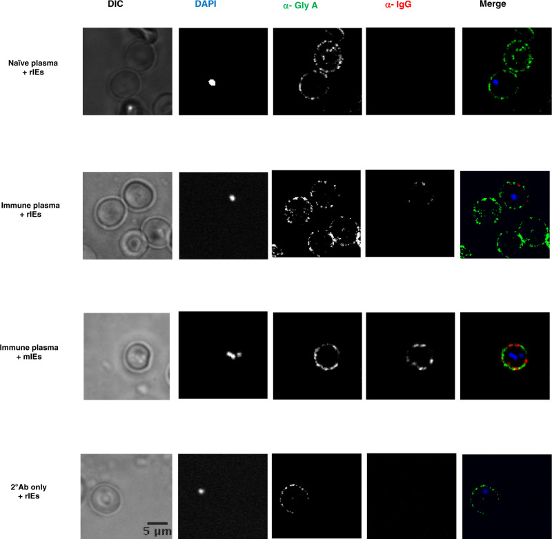
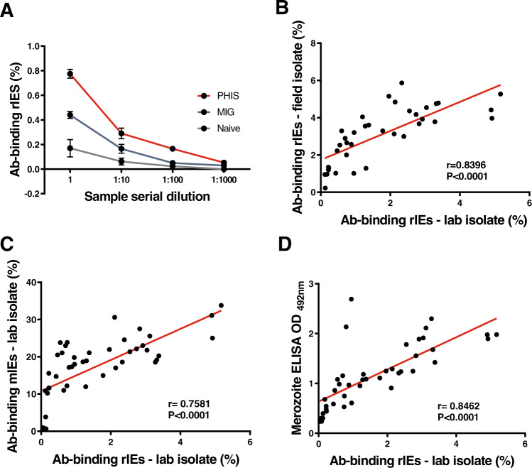
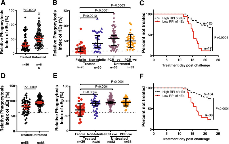
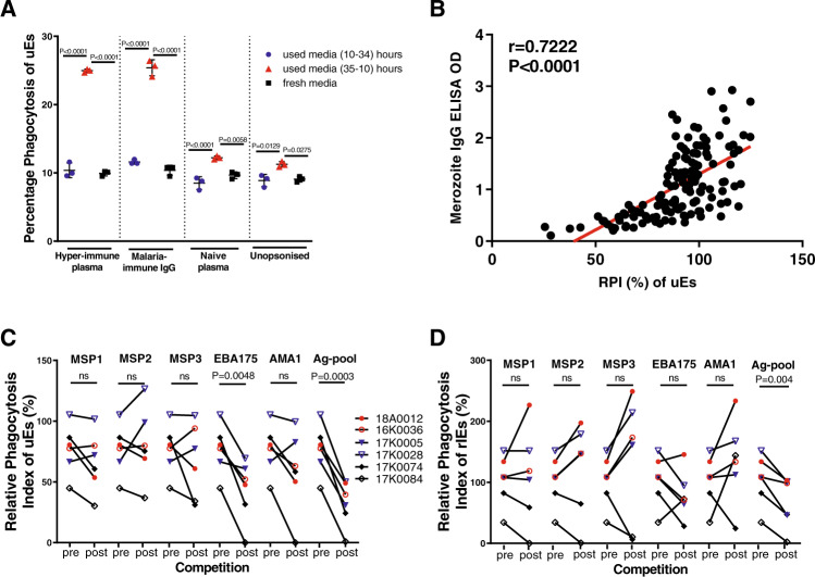
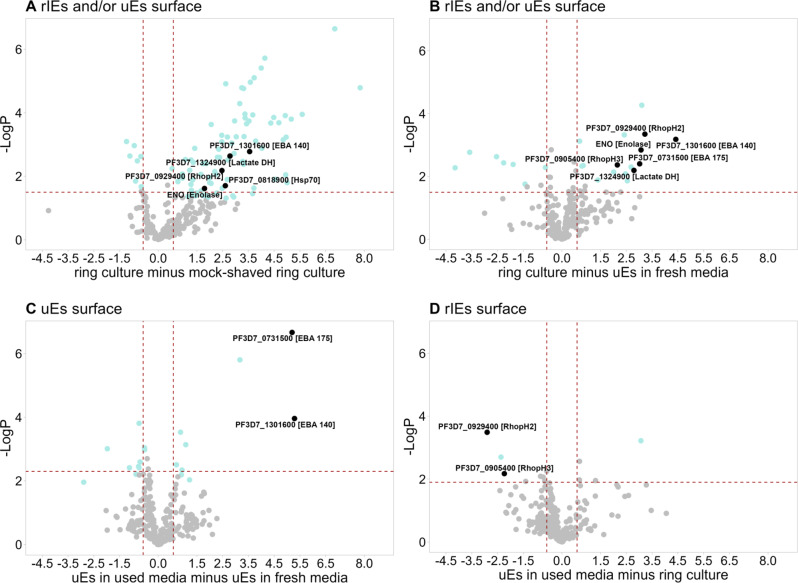
Ring-infected erythrocytes are the predominant asexual stage in the peripheral circulation but are rarely investigated in the context of acquired immunity against Plasmodium falciparum malaria. Here we compare antibody-dependent phagocytosis of ring-infected parasite cultures in samples from a controlled human malaria infection (CHMI) study (NCT02739763). Protected volunteers did not develop clinical symptoms, maintained parasitaemia below a predefined threshold of 500 parasites/μl and were not treated until the end of the study. Antibody-dependent phagocytosis of both ring-infected and uninfected erythrocytes from parasite cultures was strongly correlated with protection. A surface proteomic analysis revealed the presence of merozoite proteins including erythrocyte binding antigen-175 and -140 on ring-infected and uninfected erythrocytes, providing an additional antibody-mediated protective mechanism for their activity beyond invasion-inhibition. Competition phagocytosis assays support the hypothesis that merozoite antigens are the key mediators of this functional activity. Targeting ring-stage parasites may contribute to the control of parasitaemia and prevention of clinical malaria.


Conflict of interest statement

B.K.L.S., Y.A., P.F.B., S.L.H, E.R.J., TR. are salaried, full-time employees of Sanaria Inc., the manufacturer of Sanaria PfSPZ Challenge. Thus, all authors associated with Sanaria Inc. have potential conflicts of interest. All other authors declare no competing interests.

Figures

Fig. 1
Fig. 1. Detection of antibody binding to ring-infected erythrocytes (rIEs).
Malaria-immune plasma shows modest to low levels of binding to rIEs in comparison to mIEs in immunofluorescence assays. No binding detected with malaria-naive serum or with the secondary antibody in the absence of malaria-immune plasma. Representative data from 3 independent experiments with similar results. Source data are provided as a Source Data file.
Fig. 2
Fig. 2. Characteristics of antibodies binding to rIEs.
A IgG antibody binding to rIEs was dose-dependent in human control samples. Error bars show the mean and standard error of the mean (SEM). N = 3 biologically independent controls were analyzed in duplicate in n = 2 independent experiments. Positive controls were pooled hyperimmune serum (PHIS, red lines) and malaria-immune globulin (MIG, blue lines), while negative controls were pooled malaria-naive serum (naive, black lines). IgG antibody binding to rIEs was strongly correlated between B field and laboratory isolates, as well as with IgG binding to C mIEs and D merozoites. Each black dot in 2B-D shows the level of antibody binding in biologically independent human samples (n = 37) from the Junju cohort to rIEs from laboratory or field isolates or to merozoites. Laboratory isolate refers to the FCR-3 strain of P. falciparum. Merozoites were of the NF54 strain used in the CHMI study. Correlations from single experiments conducted with n = 2 technical replicates per sample per assay in 2B-D were tested using Spearman’s R. Source data are provided as a Source Data file.
Fig. 3
Fig. 3. Opsonic phagocytosis of ring-infected and uninfected erythrocytes predicts protection.
The relative phagocytosis index (RPI) for ring-infected erythrocytes (rIEs) A or uninfected erythrocytes (uEs) D was compared between plasma samples from volunteers that did (n = 56, closed circles) or did not (n = 86, open circles) require treatment after a malaria challenge. Data points represent a total of N = 142 biologically independent samples tested in a single experiment with two technical replicates. The red lines indicate the median and 95% confidence intervals. Dotted horizontal lines indicate the seropositivity level (mean plus three standard deviations of malaria-naive plasma) for phagocytosis. P value determined by two-tailed Mann–Whitney U test with no adjustment for multiple comparisons. Sub-group analysis for rIEs (B) and uEs (E) respectively for treated volunteers who either developed fever (febrile, n = 26, red circles) or did not (non-febrile, n = 30, blue triangles), and for untreated volunteers in whom parasites were either detected by PCR (PCR + ve, n = 53, purple triangles) or remained negative (PCR-ve, n = 33, orange diamonds). Data points represent a total of N = 142 biologically independent samples tested in a single experiment with two technical replicates. The black lines indicate the median and 95% confidence intervals. Dotted horizontal lines indicate the seropositivity level for phagocytosis. P values were determined by Kruskal–Wallis two-sided test followed by Dunn’s multiple comparison test. C & F Kaplan–Meier curves for the time to treatment for volunteers with a high (n = 125, black line) versus low (n = 17, red line) RPI against rIEs, and a high (n = 104, black line) versus low (n = 38, red line) RPI against uEs, respectively. A total of N = 142 biologically independent samples were analysed in n = 2 independent experiments with n = 2 technical replicates per experiment. P value determined by log-rank test in a Kaplan–Meier survival analysis and not adjusted for confounding variables. Source data are provided as a Source Data file.
Fig. 4
Fig. 4. Merozoite antigens are likely targets of antibody-dependent opsonic phagocytosis of ring-infected and uninfected erythrocytes.
A Phagocytosis of freshly isolated uEs that were pre-incubated with supernatants from mid-cycle parasite cultures (10 h to 34 h, blue circles), peri-invasion cultures (35 h to 10 h post invasion, red triangles), and fresh culture media (black squares). P values from a one-way ANOVA test followed by Tukey’s multiple comparison test. The graph shows the mean and error bars indicate the standard deviation. Each data point represents one of three biological replicates. B Phagocytosis of rIEs is strongly correlated with total IgG antibody binding to merozoites (n = 142). Each black dot indicates the level of IgG binding to either merozoites or RPI (%) of uEs in a total of N = 142 biologically independent samples, tested in a single experiment in duplicate. Correlation is tested using Spearmans’ R. The RPI of C uEs and D rIEs using individual plasma samples (n = 6) before (pre) and after (post) competition with individual and pooled recombinant merozoite antigens. Coloured symbols denote individual samples with high (n = 2, red), medium (n = 2, blue) and low (n = 2, black) levels of IgG against merozoites, respectively. P values were determined using a two-tailed paired t-test. Each data point represents the RPI measured once for each plasma sample with two technical replicates. RPI, relative phagocytosis index. Source data are provided as a Source Data file.
Fig. 5
Fig. 5. Volcano plots demonstrate significant enrichment of distinct merozoite proteins in specific experimental conditions.
Each panel shows the abundance of individual proteins after a comparative analysis of the following experimental conditions (x-axis): A ring cultures versus mock-shaved ring cultures, B ring cultures versus uEs in fresh media, C uEs in used media versus uEs in fresh media, and D uEs in used media versus ring cultures. Black and green dots in each panel indicate the Plasmodial and human proteins, respectively that were detected significantly more abundantly in one experimental condition compared to the other. Grey dots indicate proteins that were not detected differentially in the respective experimental conditions. For each panel, subheadings are the suggested localisation of the Plasmodium proteins shown in black. The negative Log10 p-values are displayed on the y-axis and the Log2-fold changes are displayed on the x-axis. The dotted red lines indicate the 0.05% FDR significance cut-off. Differences between pairs of treatment conditions were calculated using two-sample t-tests without correction for multiple comparisons. Four independent biological replicates were analysed for each experimental condition. Source data are uploaded onto the PRIDE database.
Fig. 6
Fig. 6. Association of phagocytosis of rIEs and uEs with high haemoglobin levels in CHMI individuals.
A Hb levels were measured once in biologically independent blood samples from volunteers (n = 142) at n = 2-time points on the day before the challenge (C-1, open squares) and the day of discharge (DOD, closed squares). Each data point represents the Hb level from independent volunteers at distinct time points. Red lines indicate the mean and 95% confidence intervals. P value determined by a two-sided Wilcoxon signed-rank t test for matched pairs. B Hb levels measured on DOD subtracted from those on C-1 for treated (n = 56, closed circles) versus non-treated (n = 86, open circles) volunteers. Each data point represents the difference in Hb levels between C-1 and DOD for each volunteer. Red lines indicate the mean and 95% confidence intervals. The dotted horizontal line indicates no difference between C-1 and DOD. The P value comparing the decrease in Hb was determined by a two-tailed Mann–Whitney U test. C Sub-group analysis for a decrease in Hb for treated volunteers who either developed fever (febrile, n = 26, red circles) or did not (non-febrile, n = 30, blue triangles), and untreated volunteers in whom parasites were either detected by PCR (PCR + ve, n = 53, purple triangles) or remained negative (PCR-ve, n = 33, orange diamonds). Each data point represents the difference in Hb levels between C-1 and DOD. Black lines indicate the mean and 95% confidence intervals. P values comparing the decrease in Hb were determined by the Kruskal–Wallis test followed by Dunn’s multiple comparison test. Source data are provided as a Source Data file.

Comment in

Similar articles

Cited by

References

    1. Cowman AF, Healer J, Marapana D, Marsh K. Malaria: biology and disease. Cell. 2016;167:610–624. doi: 10.1016/j.cell.2016.07.055. - DOI - PubMed
    1. Yazdani SS, Mukherjee P, Chauhan VS, Chitnis CE. Immune responses to asexual blood-stages of malaria parasites. Curr. Mol. Med. 2006;6:187–203. doi: 10.2174/156652406776055212. - DOI - PubMed
    1. Douki JB, et al. Adhesion of normal and Plasmodium falciparum ring-infected erythrocytes to endothelial cells and the placenta involves the rhoptry-derived ring surface protein-2. Blood. 2003;101:5025–5032. doi: 10.1182/blood-2002-12-3710. - DOI - PubMed
    1. Pouvelle B, Buffet PA, Lepolard C, Scherf A, Gysin J. Cytoadhesion of Plasmodium falciparum ring-stage-infected erythrocytes. Nat. Med. 2000;6:1264–1268. doi: 10.1038/81374. - DOI - PubMed
    1. Buffet PA, et al. Ex vivo perfusion of human spleens maintains clearing and processing functions. Blood. 2006;107:3745–3752. doi: 10.1182/blood-2005-10-4094. - DOI - PubMed

Publication types

Associated data