. 2022 Jul 14;12(1):12025.
doi: 10.1038/s41598-022-14049-5.

Chromosomal phase improves aneuploidy detection in non-invasive prenatal testing at low fetal DNA fractions

Affiliations

Chromosomal phase improves aneuploidy detection in non-invasive prenatal testing at low fetal DNA fractions

Giulio Genovese et al. Sci Rep. .

Abstract

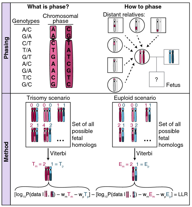
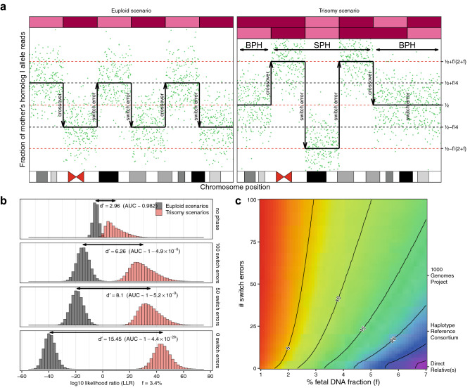
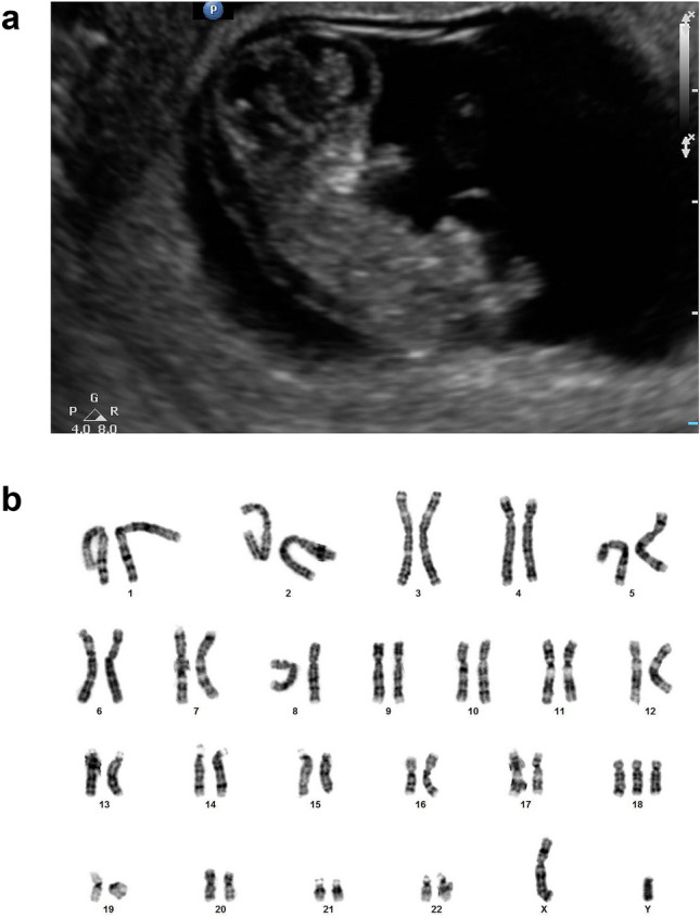
Non-invasive prenatal testing (NIPT) to detect fetal aneuploidy by sequencing the cell-free DNA (cfDNA) in maternal plasma is being broadly adopted. To detect fetal aneuploidies from maternal plasma, where fetal DNA is mixed with far-larger amounts of maternal DNA, NIPT requires a minimum fraction of the circulating cfDNA to be of placental origin, a level which is usually attained beginning at 10 weeks gestational age. We present an approach that leverages the arrangement of alleles along homologous chromosomes-also known as chromosomal phase-to make NIPT analyses more conclusive. We validate our approach with in silico simulations, then re-analyze data from a pregnant mother who, due to a fetal DNA fraction of 3.4%, received an inconclusive aneuploidy determination through NIPT. We find that the presence of a trisomy 18 fetus can be conclusively inferred from the patient's same molecular data when chromosomal phase is incorporated into the analysis. Key to the effectiveness of our approach is the ability of homologous chromosomes to act as natural controls for each other and the ability of chromosomal phase to integrate subtle quantitative signals across very many sequence variants. These results show that chromosomal phase increases the sensitivity of a common laboratory test, an idea that could also advance cfDNA analyses for cancer detection.


Conflict of interest statement

Giulio Genovese, Po-Ru Loh, and Steven A. Mccarroll declare competing interests: patent application PCT/WO2019/079493 has been filed on the mosaic chromosomal alterations detection method used in this work. This does not restrict the non-commercial use of the method described in this article. The other authors have no competing interests.

Figures

Figure 1
Figure 1
Schematic representation of the role of chromosomal phase in the computation of the discrimination statistic Graphical representation of how chromosomal phase is inferred through relatives on the top and how the space of possible fetal inheritance configurations is explored on the bottom to identify, through the Viterbi algorithm, the two most likely configurations for both the trisomy and the euploid scenario. A log10 likelihood ratio (LLR) discrimination statistic based on the likelihood of the data and weighed by the number of crossovers and phase switch errors needed to explain the fetal genome is then computed. The weight applied to the number of crossovers and switch errors is akin to assigning a prior over the space of possible fetal inheritance configurations that concentrates the probability towards configurations that can be explained with fewer switches starting from parental homologs.
Figure 2
Figure 2
Simulation of log10 likelihood ratio (LLR) discrimination statistics. (a) Simulated allelic fractions from the maternal plasma of a pregnant woman with crossovers and switch errors. The top bars represent the simulated maternal homologs inherited by the fetus, with magenta and red representing, respectively, mother’s homolog I and mother’s homolog II. Notice that, in the trisomy scenario, switch errors don’t change the expected proportion of alleles when occurring in BPH segments. (b) Simulated log10 likelihood ratio (LLR) discrimination statistics from simulations with a fetal DNA fraction specified as f = 3.4%, and an average sampling of 2000 sequence fragments at 1500 loci heterozygous for the mother, for both trisomy and euploid scenarios. Sensitivity index d’ between the LLR discrimination statistics for the two models is displayed together with the AUC estimated as if the two LLR discrimination statistics were normally distributed. As the number of switch errors decreases, the ability of the LLR discrimination statistic to distinguish the euploid and trisomy scenarios increases. (c) Sensitivity index d’ between LLR discrimination statistics for allelic read counts simulated by trisomy scenarios and those simulated by euploid scenarios as a function of fetal DNA fraction and the number of switch errors. For reference, a representative chromosomal phase accuracy using different chromosomal phasing approaches is reported on the right. Contour lines follow parameters sets with the same sensitivity index d’, indicating scenarios with approximately equivalent power to distinguish euploid and trisomy scenarios.
Figure 3
Figure 3
Fetal ultrasound and karyotype. (a) Ultrasound image of a trisomy 18 fetus at 11 weeks GA showing potential neck thickening and a crown-rump length of 33 mm. (b) Fetal karyotype. Abnormal 47,XY, + 18 male chromosome karyotype with an extra chromosome 18 observed in mitotic cells obtained from examination of products of conception.
Figure 4
Figure 4
SNP-based targeted sequencing data without chromosomal phase. Graphical representation of sequencing data from the SNP-based targeted sequencing of maternal plasma obtained from the pregnant mother at 11 weeks GA with a fetal DNA fraction estimated as f = 3.4%. Points correspond to the fraction of alternate (A) allele read counts as a fraction of the overall number of reads at SNP loci sampled by more than 200 sequence fragments. At loci for which the mother is homozygous, the fetal genotype can be inferred with high accuracy. Black and red dotted lines represent the expected fractions of A alleles for different combinations of maternal and fetal genotypes in the case of, respectively, a euploid and trisomic fetus. The lack of SNP loci at fractions for which the mother is expected to be homozygous and the fetus is expected to be heterozygous along the chromosome X homologs is indicative of a male fetus.
Figure 5
Figure 5
SNP-based targeted sequencing data with chromosomal phase. (a) Pedigree with parents and grandparents of the trisomy 18 fetus. MPSS data of DNA from saliva were available for the parents and microarray genotype data of DNA from saliva were available for the grandparents. Data from the SNP-based targeted sequencing and MPSS of maternal plasma using the Illumina Nextseq 500 platform were available at, respectively, 11 weeks and 15 weeks GA. (b) Schematic representation of the formation of a trisomic zygote through missegregation of chromosomes during maternal meiosis II. (c) Graphical representation of sequence data at loci heterozygous for the mother from the SNP-based targeted sequencing of maternal plasma at 11 weeks GA with a fetal DNA fraction estimated as f = 3.4%. Each green point corresponds to the fraction of the mother’s maternal allele reads at any of the 13,926 SNP loci that are consistent with heterozygous genotypes for the mother and were covered by more than 200 sequence fragments. The two black dotted lines represent the expected fractions of the mother’s maternal alleles in the case of a euploid fetus, and the three red dotted lines represent the expected fractions in the case of a fetus with trisomy of maternal origin. The blue line is a centered rolling mean across 200 consecutive heterozygous SNPs. The top bars represent the inferred inherited homologs of the fetus, with magenta, red, cyan, and blue colors representing, respectively, mother’s maternal, mother’s paternal, father’s maternal, and father’s paternal homologs. Chromosome 18, with three fetal homologs inferred, is highlighted in red. It is important to note that the algorithm to infer the inherited homolog segments takes also into account information about the homologs transmitted from the father of the fetus and allelic fractions at SNP loci consistent with homozygous genotype for the mother and which are not displayed in this figure and that the paternal homologs of the fetus further adds to the sampling noise at loci heterozygous for the mother.

References

    1. Lo YM, et al. Presence of fetal DNA in maternal plasma and serum. Lancet Lond. Engl. 1997;350:485–487. doi: 10.1016/S0140-6736(97)02174-0. - DOI - PubMed
    1. Bianchi DW, et al. DNA sequencing versus standard prenatal aneuploidy screening. N. Engl. J. Med. 2014;370:799–808. doi: 10.1056/NEJMoa1311037. - DOI - PubMed
    1. Pergament E, et al. Single-nucleotide polymorphism-based noninvasive prenatal screening in a high-risk and low-risk cohort. Obstet. Gynecol. 2014;124:210–218. doi: 10.1097/AOG.0000000000000363. - DOI - PMC - PubMed
    1. Porreco RP, et al. Noninvasive prenatal screening for fetal trisomies 21, 18, 13 and the common sex chromosome aneuploidies from maternal blood using massively parallel genomic sequencing of DNA. Am. J. Obstet. Gynecol. 2014;211(365):e1–12. - PubMed
    1. Zhang, H. et al. Non-invasive prenatal testing for trisomies 21, 18 and 13: clinical experience from 146,958 pregnancies. Ultrasound Obstet. Gynecol. Off. J. Int. Soc. Ultrasound Obstet. Gynecol.45, 530–538 (2015). - PubMed

Publication types