. 2022 Jul 14;13(1):4085.
doi: 10.1038/s41467-022-31819-x.

Auxotrophic and prototrophic conditional genetic networks reveal the rewiring of transcription factors in Escherichia coli

Affiliations

Auxotrophic and prototrophic conditional genetic networks reveal the rewiring of transcription factors in Escherichia coli

Alla Gagarinova et al. Nat Commun. .

Abstract

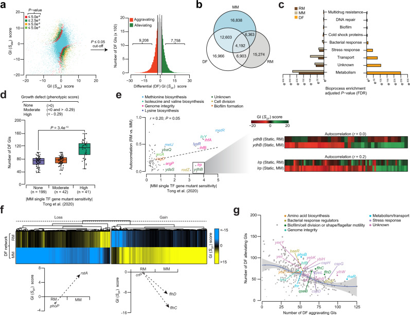
Bacterial transcription factors (TFs) are widely studied in Escherichia coli. Yet it remains unclear how individual genes in the underlying pathways of TF machinery operate together during environmental challenge. Here, we address this by applying an unbiased, quantitative synthetic genetic interaction (GI) approach to measure pairwise GIs among all TF genes in E. coli under auxotrophic (rich medium) and prototrophic (minimal medium) static growth conditions. The resulting static and differential GI networks reveal condition-dependent GIs, widespread changes among TF genes in metabolism, and new roles for uncharacterized TFs (yjdC, yneJ, ydiP) as regulators of cell division, putrescine utilization pathway, and cold shock adaptation. Pan-bacterial conservation suggests TF genes with GIs are co-conserved in evolution. Together, our results illuminate the global organization of E. coli TFs, and remodeling of genetic backup systems for TFs under environmental change, which is essential for controlling the bacterial transcriptional regulatory circuits.


Conflict of interest statement

The authors declare no competing interests

Figures

Fig. 1
Fig. 1. TF gene targets and static and differential GI screening pipeline.
a Schematic of the conjugation, double mutant generation in RM and MM growth conditions, colony imaging, mutant strain growth rate measurements (n = 8 replicate gene pairs per donor) using multiplicative and Gaussian process models, and construction of static and differential genetic networks using the significant GI scores derived from respective growth conditions. b, c TF genes screened as donors and recipients (b) to generate static and differential networks, and their assignment (c) into 13 broadly representative bioprocesses. Source Data are provided as a Source Data file.
Fig. 2
Fig. 2. Epistatic patterns/connectivity of TFs in static RM network.
a Enrichment (P-value by one-tailed hypergeometric test, and adjusted using the Benjamini-Hochberg FDR correction) of crosstalk among bioprocesses (n = 254 of 1431 process pairs tested) with distinct aggravating or alleviating GI patterns in RM is shown along with illustrative examples (see also Supplementary Fig. 2a). b Representative regulatory modules involving TF gene pairs as regulators, activators, or repressors of gene expression, and their respective epistatic relationships are shown. Since StpA and H-NS have overlapping and distinct functions, we predict (indicated in question mark) that like hns, stpA may repress or regulate acid resistance (AR) genes based on the alleviating GI patterns with AR targets. c, d Enrichment of aggravating (P = 1.8 e−7) or alleviating (P = 3.2 e−8) GIs (c; from left to right, n = 17 and 27 gene pairs; P-value by hypergeometric test), and epistatic connectivity (d; from left to right, n = 27 and 35 genes) between TF genes that function as co-repressors or co-activators (P = 1.0 e−3 by two-sided Wilcoxon signed-rank test). Box plot (d) from left to right is shown with maximum (0.055 and 0.044), minimum (0.021 and 0.013), quartile 1 (Q1; 0.029 and 0.022), Q2 (0.037 and 0.027) and Q3 (0.047 and 0.035) values. e, f GI frequency (e; from left to right, n = 182 and 182 genes; P = 2.2 e−16 by two-sided Wilcoxon signed-rank test), and ratio of aggravating to alleviating GIs (f; from left to right, n = 182 and 182 genes; P = 1.4 e−5 by two-sided Wilcoxon signed-rank test) observed between local TFs compared to local with global TF gene pairs. Box plot (e) from left to right is shown with maximum (0.009 and 0.010), minimum (0.0015 and 0.00), Q1 (0.0041 and 0.003), Q2 (0.005 and 0.0055) and Q3 (0.0058 and 0.008) values. Source data are provided as a Source Data file.
Fig. 3
Fig. 3. Orphan TF epistatic connections in static RM network.
a Frequency of orphan TF interactions with annotated TF genes in the indicated bioprocesses. b Sub-network of two orphan TF genes (yddM, ygfI) interacting with stress response regulators, and their correlated GI profiles (n = 300 gene pairs). c Distribution of orphan TF gene pairs (n = 1913) interacting with one or more global regulators. d Target global regulators, and their GI frequency with orphan TF genes. e Correlated GI profiles between yjdC and dicA or dicC in RM and MM (from top to bottom, n = 300 gene pairs for RM and MM, respectively). f, g Growth rate (f; n = 12 biologically independent experiments; P = 4.0 e−9 by Student’s two-sided t-test) at 32 °C with OD600 measured at 24 h, and representative cell morphology micrographs (T = 4 h after reaching OD = 0.5–0.6) along with cell length measurements (g; n ≥ 25 cells over five biologically independent experiments; P = 6.4 e−31 and 6.1 e−30 by Student’s two-sided t-test) of essential dicA hypomorph (*) or dicC mutant and yjdC double or single mutants, or overexpression of yjdC in wild-type (WT), dicC mutant or dicA hypomorph in RM. Scale bar represents 10 μm. h Fold enrichment of YjdC binding to dicABC genes by ChIP-qPCR in RM and MM (n = 3 biologically independent experiments) was analyzed by comparing chromosomal YjdC FLAG3-tag to untagged control strain, and normalized to a negative control (i.e., primers designed away from YjdC binding site of dic locus was used to amplify from YjdC-FLAG3). i Transcript levels of yjdC and dic genes relative to WT measured by qRT-PCR (n = 3 biologically independent experiments) in the strains indicated. Significance in panels h and i (*P ≤ 0.05, **P ≤ 0.01, ***P ≤ 0.001, **** P ≤ 0.0001) are calculated using Student’s two-sided t-test. j Model illustrating the potential role of YjdC in cell division. Data (fi) are presented as mean ± standard deviation from the indicated number of independent samples. Source data are provided as a Source Data file.
Fig. 4
Fig. 4. YneJ regulates the components of putrescine pathway and efflux pump.
a Growth rate of indicated strains at 32 °C with OD600 measured (n = 10 biologically independent experiments; P = 4.4 e−10 by Student’s two-sided t-test) at 24 h in RM. b Location of yneJ near the σE promotor region of yneI operon, comprising sad-glsB-yneG genes, with DNA binding site (highlighted in green) predicted within the sad-yneJ intergenic region based on conservation analysis. c Genes in putrescine utilization and Gln-Glu pathways. d Transcript levels of indicated genes in a wild-type (WT) BW25113 strain grown in M9 media with different carbon sources (0.5% Gln, Glutamine; 0.5% Glu, Glutamate; 0.4% Putr, Putrescine; 1% Try, Tryptone) relative to 0.1% M9-glucose (n = 3 biologically independent experiments). e Fold enrichment of YneJ binding site in sad-yneJ or acrA-acrR intergenic region measured by ChIP-qPCR (n = 3 biologically independent experiments) in M9-putrescine, tryptone and glucose media. ChIP enrichment in a chromosomal YneJ FLAG3-tag normalized to a negative control (i.e., primers designed away from YneJ binding site of sad or acrR was used to amplify from YneJ-FLAG3). f YneJ binding to sad-yneJ site by fluorescence polarization (n = 4 biologically independent experiments) is shown at increasing concentration of YneJ recombinant protein with 10 nM fluorescence-labeled double-stranded DNA (i.e., sad-yneJ consensus motif or promoter region of leuO gene that serve as negative control), and in the presence or absence of Gln (2 mM). Significance (****P = 1.7 e−9) calculated using Student’s two-sided t-test between sad-yneJ consensus motif with Gln vs. without Gln at 1 µM concentration of YneJ protein. g Transcript levels of target genes measured by qRT-PCR (n = 3 biologically independent experiments) in yneJ mutant or complemented (+) strains vs. WT grown in M9-tryptone. h Distribution of correlation coefficient profiles among TFs, with emphasis on yneJ-acrR GI profile (n = 300 gene pairs) in RM and MM. i Growth rates of WT and indicated mutant or complemented (+) strains in M9 media containing tryptone, with or without SDS (n = 12 biologically independent experiments; P = 6.4 e−17 by Student’s two-sided t-test) at 32 °C over 24 h. j Changes in transcript levels of target genes in yneJ mutant or complemented (+) strain vs. WT grown in M9-tryptone (n = 3 biologically independent experiments). k Model illustrating YneJ role in the regulation of Puu and efflux pump. Data (a, dg, i, j) are presented as mean ± standard deviation from the indicated number of independent samples. Significance in panels d, e, g and j (*P ≤ 0.05, **P ≤ 0.01, ***P ≤ 0.001, ****P ≤ 0.0001) calculated by Student’s two-sided t-test. Source data are provided as a Source Data file.
Fig. 5
Fig. 5. Differential networks reveal new TF gene functions.
a Scatterplot (left) of GI scores of RM vs. MM static networks at various P-value-based cut-offs (≤ 5.0 e−2, 2.5 e−2, 5.0 e−3, 5.0 e−4) after Z-score transformation. Histogram (right) of differential GI scores of the Z-score transformed P-value filtered at a significance level of 0.05 with tails representing significant epistatic interactions. b Venn showing the overlap of significant GIs in static and differential networks. c Enriched TF bioprocesses (P-value significance by hypergeometric test, and adjusted using the Benjamini-Hochberg FDR correction) in static and differential networks. d, e Distribution of differential GIs (d; from left to right, n = 199, 42, and 41) or correlation of GI profiles (e) for each query TF gene between RM and MM networks (i.e., Autocorrelation) is plotted against single TF gene mutant growth fitness sensitivity in MM from a recent phenotypic genetic screen. Autocorrelation profiles for two representative query TF genes (ydhB, lrp) are shown (e); P = 3.4 e−10 by two-sided Wilcoxon signed-rank test (d) and P = 0.05 by t-distribution for Pearson’s correlation coefficient (e). Box plot (d) from left to right is shown with maximum (100, 102, and 159), minimum (39, 44, and 51), quartile 1 (Q1; 68, 72 and 93), Q2 (79, 80, and 118) and Q3 (82, 83, and 123) values. f Hierarchical clustering of GI patterns showing gene pairs with gain or loss of GI in RM or MM. Illustrative examples of two TF gene pair profiles are shown. g Differential GI hub TF genes in the indicated processes is plotted against the number of aggravating vs. alleviating differential GIs. Source data are provided as a Source Data file.
Fig. 6
Fig. 6. YdiP regulates cold shock proteins at low temperature.
a Significant (P = 5.7 e−7 by Student’s two-sided t-test) differential GIs of ydiP with other TF gene mutants in RM and MM. b, c GI patterns (b) and correlation (c) profile of ydiP in static and differential networks with genes involved in cold shock adaptation. d YdiP interaction with indicated cold shock proteins in different growth and temperature conditions by affinity purification and mass spectrometry (n = 3 biologically independent experiments). e YdiP steady-state level in RM and at different temperatures over time. Band intensities normalized to E. coli Hsp60 loading control (LC; 1:15000 dilution). Immunoblot shown is representative of 2 independent biological experiments. Molecular masses (kDa) of marker proteins are indicated. f Transcript levels of ydiP measured in wild-type (WT) and ydiP mutant strain background in RM and MM at 15 °C are shown as fold change (n = 3 biologically independent experiments; P = 1.1 e−3 by Student’s two-sided t-test) after normalizing to respective strains and growth conditions at 37 °C. g Representative cell morphology micrographs of ydiP and csp single and double mutants, as well as overexpression of ydiP (indicated with ‘+’) in ydiP mutant strain in RM at 15 °C are shown along with cell length measurements (n ≥ 25 cells over five biologically independent experiments; P = 1.8 e−28 by Student’s two-sided t-test). Scale bar, 10 µm. h Mapped reads from YdiP ChIP-seq is plotted against the position of indicated genes in genomic region, including YdiP binding to its putative cspE target in RM and MM at 15 °C. Positive value indicate plus (or sense) strand, and negative value specify minus (or antisense) strand. Fold enrichment (shown as a zoom-in; n = 3 biologically independent experiments) of YdiP binding site to cspE using a chromosomal YdiP FLAG3-tagged strain was compared to an untagged strain after normalizing to a negative control (i.e., primers designed away from YdiP binding site of cspE was amplified from YdiP- FLAG3) in different growth and temperature conditions. Significance (P = 3.4 e−4 or P = 4.5 e−5) by Student’s two-sided t-test between the YdiP FLAG3-tagged strain grown in RM or MM at 15 °C and 37 °C. i Transcript levels of csp genes measured in WT and ydiP mutant in RM and MM at 15 °C are shown as fold change (n = 3 biologically independent experiments) after normalizing to respective strains and growth conditions at 37 °C. Significance (*P ≤ 0.05, **P ≤ 0.01, ***P ≤ 0.001, ****P ≤ 0.0001) by Student’s two-sided t-test. j Model illustrating YdiP as an activator and repressor of various cold shock proteins in RM and MM growth conditions. Data (fi) are presented as mean ± standard deviation from the indicated number of independent samples. Source data are provided as a Source Data file.

Similar articles

Cited by

References

    1. Mejia-Almonte C, et al. Redefining fundamental concepts of transcription initiation in bacteria. Nat. Rev. Genet. 2020;21:699–714. doi: 10.1038/s41576-020-0254-8. - DOI - PMC - PubMed
    1. Gao Y, et al. Unraveling the functions of uncharacterized transcription factors in Escherichia coli using ChIP-exo. Nucleic Acids Res. 2021;49:9696–9710. doi: 10.1093/nar/gkab735. - DOI - PMC - PubMed
    1. Gao Y, et al. Systematic discovery of uncharacterized transcription factors in Escherichia coli K-12 MG1655. Nucleic Acids Res. 2018;46:10682–10696. - PMC - PubMed
    1. Martinez-Antonio A, Collado-Vides J. Identifying global regulators in transcriptional regulatory networks in bacteria. Curr. Opin. Microbiol. 2003;6:482–489. doi: 10.1016/j.mib.2003.09.002. - DOI - PubMed
    1. Eisen MB, Spellman PT, Brown PO, Botstein D. Cluster analysis and display of genome-wide expression patterns. Proc. Natl. Acad. Sci. USA. 1998;95:14863–14868. doi: 10.1073/pnas.95.25.14863. - DOI - PMC - PubMed

Publication types

MeSH terms

Substances

Grants and funding

LinkOut - more resources