

Tissue volume estimation and age prediction using rapid structural brain scans
- PMID: 35835813
- PMCID: PMC9283414
- DOI: 10.1038/s41598-022-14904-5
Tissue volume estimation and age prediction using rapid structural brain scans
Abstract
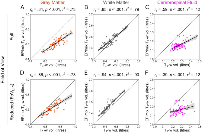
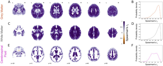
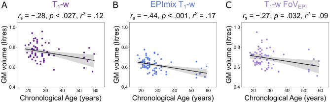
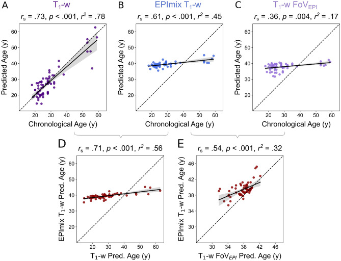
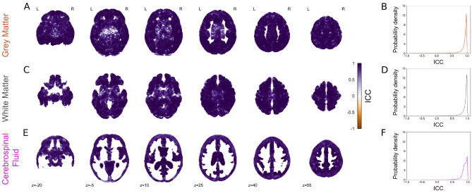
The multicontrast EPImix sequence generates six contrasts, including a T1-weighted scan, in ~1 min. EPImix shows comparable diagnostic performance to conventional scans under qualitative clinical evaluation, and similarities in simple quantitative measures including contrast intensity. However, EPImix scans have not yet been compared to standard MRI scans using established quantitative measures. In this study, we compared conventional and EPImix-derived T1-weighted scans of 64 healthy participants using tissue volume estimates and predicted brain-age. All scans were pre-processed using the SPM12 DARTEL pipeline, generating measures of grey matter, white matter and cerebrospinal fluid volume. Brain-age was predicted using brainageR, a Gaussian Processes Regression model previously trained on a large sample of standard T1-weighted scans. Estimates of both global and voxel-wise tissue volume showed significantly similar results between standard and EPImix-derived T1-weighted scans. Brain-age estimates from both sequences were significantly correlated, although EPImix T1-weighted scans showed a systematic offset in predictions of chronological age. Supplementary analyses suggest that this is likely caused by the reduced field of view of EPImix scans, and the use of a brain-age model trained using conventional T1-weighted scans. However, this systematic error can be corrected using additional regression of T1-predicted brain-age onto EPImix-predicted brain-age. Finally, retest EPImix scans acquired for 10 participants demonstrated high test-retest reliability in all evaluated quantitative measurements. Quantitative analysis of EPImix scans has potential to reduce scanning time, increasing participant comfort and reducing cost, as well as to support automation of scanning, utilising active learning for faster and individually-tailored (neuro)imaging.
© 2022. The Author(s).
Conflict of interest statement
The authors declare no competing interests.
Figures
References
-
- Delgado, A. F. et al. Diagnostic performance of a new multicontrast one-minute full brain exam ( EPIMix ) in neuroradiology: A prospective study. J. Magn. Reson. Imaging, 1–10 (2019). - PubMed
-
- Mekle, R., Wu, E. X., Meckel, S., Wetzel, S. G. & Scheffler, K. Combo acquisitions: Balancing scan time reduction and image quality. Magn. Reson. Med.55, 1093–1105. issn: 0740-3194 (2006). - PubMed
-
- Andre, J. B. et al. Toward quantifying the prevalence, severity, and cost associated with patient motion during clinical MR examinations. J. Am. Coll. Radiol.12, 689–695. issn: 1546- 1440 (2015). - PubMed
Publication types
MeSH terms
Associated data
Grants and funding
LinkOut - more resources
Full Text Sources
Medical
