Mouse oocytes carrying metacentric Robertsonian chromosomes have fewer crossover sites and higher aneuploidy rates than oocytes carrying acrocentric chromosomes alone
- PMID: 35835815
- PMCID: PMC9283534
- DOI: 10.1038/s41598-022-16175-6
Mouse oocytes carrying metacentric Robertsonian chromosomes have fewer crossover sites and higher aneuploidy rates than oocytes carrying acrocentric chromosomes alone
Abstract
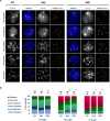
Meiotic homologous recombination during fetal development dictates proper chromosome segregation in adult mammalian oocytes. Successful homologous synapsis and recombination during Meiotic Prophase I (MPI) depends on telomere-led chromosome movement along the nuclear envelope. In mice, all chromosomes are acrocentric, while other mammalian species carry a mixture of acrocentric and metacentric chromosomes. Such differences in telomeric structures may explain the exceptionally low aneuploidy rates in mice. Here, we tested whether the presence of metacentric chromosomes carrying Robertsonian translocations (RbT) affects the rate of homologous recombination or aneuploidy. We found a delay in MPI progression in RbT-carrier vs. wild-type (WT) fetal ovaries. Furthermore, resolution of distal telomere clusters, associated with synapsis initiation, was delayed and centromeric telomere clusters persisted until later MPI substages in RbT-carrier oocytes compared to WT oocytes. When chromosomes fully synapsed, higher percentages of RbT-carrier oocytes harbored at least one chromosome pair lacking MLH1 foci, which indicate crossover sites, compared to WT oocytes. Aneuploidy rates in ovulated eggs were also higher in RbT-carrier females than in WT females. In conclusion, the presence of metacentric chromosomes among acrocentric chromosomes in mouse oocytes delays MPI progression and reduces the efficiency of homologous crossover, resulting in a higher frequency of aneuploidy.
© 2022. The Author(s).
Conflict of interest statement
The authors declare no competing interests.
Figures


Similar articles
-
Two telomeric ends of acrocentric chromosome play distinct roles in homologous chromosome synapsis in the fetal mouse oocyte.Chromosoma. 2021 Mar;130(1):41-52. doi: 10.1007/s00412-021-00752-1. Epub 2021 Jan 25. Chromosoma. 2021. PMID: 33492414
-
SIRT7 promotes chromosome synapsis during prophase I of female meiosis.Chromosoma. 2019 Sep;128(3):369-383. doi: 10.1007/s00412-019-00713-9. Epub 2019 Jun 29. Chromosoma. 2019. PMID: 31256246 Free PMC article.
-
Nondisjunction, disturbances in spindle structure, and characteristics of chromosome alignment in maturing oocytes of mice heterozygous for Robertsonian translocations.Cytogenet Cell Genet. 1990;54(1-2):47-54. doi: 10.1159/000132953. Cytogenet Cell Genet. 1990. PMID: 2249474
-
Regulation of Meiotic Prophase One in Mammalian Oocytes.Front Cell Dev Biol. 2021 May 20;9:667306. doi: 10.3389/fcell.2021.667306. eCollection 2021. Front Cell Dev Biol. 2021. PMID: 34095134 Free PMC article. Review.
-
A bouquet of chromosomes.J Cell Sci. 2004 Aug 15;117(Pt 18):4025-32. doi: 10.1242/jcs.01363. J Cell Sci. 2004. PMID: 15316078 Review.
Cited by
-
Fertility Cost (or Sometimes a Lack of It) in Relation to Heterozygosity for Robertsonian Rearrangements in Mammals: A Review.Cytogenet Genome Res. 2025 May 30:1-35. doi: 10.1159/000546385. Online ahead of print. Cytogenet Genome Res. 2025. PMID: 40451160 Free PMC article. Review.
References
-
- Kuliev A, Zlatopolsky Z, Kirillova I, Spivakova J, Cieslak Janzen J. Meiosis errors in over 20,000 oocytes studied in the practice of preimplantation aneuploidy testing. RBM Online. 2011;22:2–8. - PubMed
-
- Kalmbach KH, Antunes DMF, Kohlrausch F, Keefe DL. Seminars in Reproductive Medicine. Thieme Medical Publishers; 2022. pp. 389–395. - PubMed
Publication types
MeSH terms
Grants and funding
LinkOut - more resources
Full Text Sources
Molecular Biology Databases

