. 2022 Jul 14;5(1):704.
doi: 10.1038/s42003-022-03656-7.

Conformational changes in the catalytic region are responsible for heat-induced activation of hyperthermophilic homoserine dehydrogenase

Affiliations

Conformational changes in the catalytic region are responsible for heat-induced activation of hyperthermophilic homoserine dehydrogenase

Tatsuya Kubota et al. Commun Biol. .

Abstract

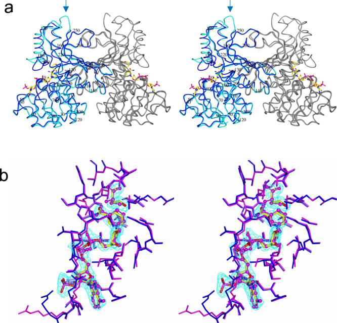
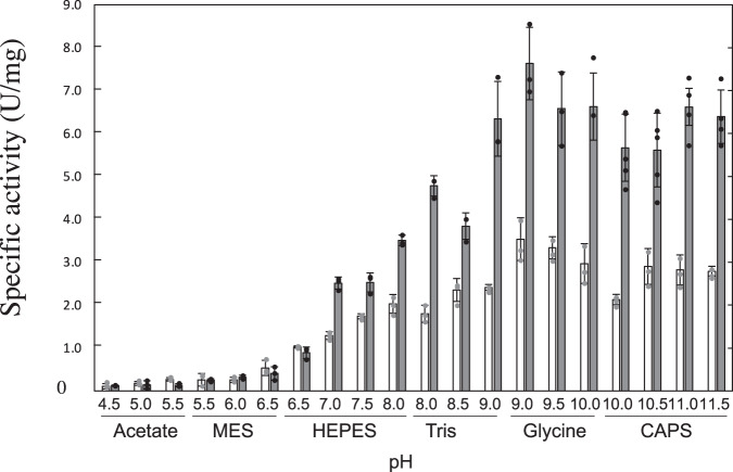
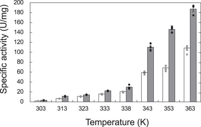
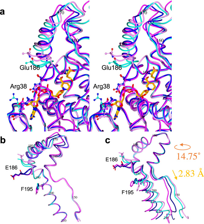
When overexpressed as an immature enzyme in the mesophilic bacterium Escherichia coli, recombinant homoserine dehydrogenase from the hyperthermophilic archaeon Sulfurisphaera tokodaii (StHSD) was markedly activated by heat treatment. Both the apo- and holo-forms of the immature enzyme were successively crystallized, and the two structures were determined. Comparison among the structures of the immature enzyme and previously reported structures of mature enzymes revealed that a conformational change in a flexible part (residues 160-190) of the enzyme, which encloses substrates within the substrate-binding pocket, is smaller in the immature enzyme. The immature enzyme, but not the mature enzyme, formed a complex that included NADP+, despite its absence during crystallization. This indicates that the opening to the substrate-binding pocket in the immature enzyme is not sufficient for substrate-binding, efficient catalytic turnover or release of NADP+. Thus, specific conformational changes within the catalytic region appear to be responsible for heat-induced activation.


Conflict of interest statement

The authors declare no competing interests.

Figures

Fig. 1
Fig. 1. The IM and IM/NADP/BU structures in their physiological dimeric form.
a The IM and IM/NADP/BU structures are shown in cyan and blue, respectively, with the other subunit in gray. NADP+ atoms are shown as cylinders. The arrow indicates a disordered part (227–231 aa). b The substrate-binding pockets of the IM/NADP/BU and M/NAD/Cys structures are shown in blue and pink, respectively. NADP+ and BU are shown as cylinders, while NAD+ and Cys are shown as ball and stick. The 2Fo–Fc electron density in the map is contoured at the +1.0 sigma level.
Fig. 2
Fig. 2. Effect of pH on enzyme activity.
The activity of the immature (white bar) and mature enzymes (gray bar) were assayed in 100 mM acetate (pHs 4.5, 5,0 and 5.5), MES (pHs 5.5, 6.0 and 6.5), HEPES (pHs 6.5, 7.0, 7.5 and 8.0), Tris (pHs 8.0, 8.5 and 9.0), Glycine (pHs 9.0, 9.5 and 10.0) or CAPS (pHs 10.0, 10.5, 11.0 and 11.5) at 303 K. The length of the error bars represents the standard deviation.
Fig. 3
Fig. 3. Effect of temperature on enzyme activity.
The activity of the immature (white bar) and mature enzymes (gray bar) were assayed in 100 mM tris (pH 8.0) at various temperatures. The length of the error bars represents the standard deviation.
Fig. 4
Fig. 4. Conformational change induced by heat-treatment.
Differences between the backbone atoms of the M and IM (a) and M/NAD/Cys and IM/NADP/BU (b) structures are superimposed. The arrows and numbers indicate the residues mentioned in the text. The flexible (160–190 aa) and lid part (181–190 aa) are highlighted by two-way arrow. The helix structure (194–210 aa) described in Fig. 6 is marked with an asterisk.
Fig. 5
Fig. 5. Conformational change induced by ligand binding.
Differences between the backbone atoms of the IM and IM/NADP/BU (a) and M and M/NAD/Cys (b) structures are superimposed. The arrows and numbers indicate the residues mentioned in the text. The a-helix and b-sheet structures are shown in arrows and white boxes, respectively. The flexible (160–190 aa) and lid part (181–190 aa) are shown by two-way arrow. The helix structure (194–210 aa) described in Fig. 6 is marked with an asterisk.
Fig. 6
Fig. 6. Comparison of substrate-binding pockets.
a The substrate-binding pockets within the IM (cyan), IM/NADP/BU (blue), M (pink) and M/NAD/Cys (magenta) structures are shown. The atoms of Glu186 and Arg38 are shown as ball and stick. The substrates and substrate analogs of the IM/NADP/BU (yellow) and M/NAD/Cys (orange) structures are shown as cylinders. Side-chain atoms of Glu186 within the IM and M structures are disordered. The backbone atoms of the helix structure (194–210 aa) (b) or the rigid part (residues 160–180) (c) of the IM (cyan), IM/NADP/BU (blue), M (pink) and M/NAD/Cys (magenta) structures are superposed. Superposition of the rigid parts of the IM and M structures (b) requires 14.75˚ rotation and 2.83 Å displacement. The side chains of residues E186 and F195 are shown as cylinders.
Fig. 7
Fig. 7. Stereo-view of a part of the catalytic region.
Hydrogen bonds within the substrate-binding pocket of the IM/NADP/BU structure are shown. The 2Fo–Fc electron density in the map is contoured at the +1.0 sigma level.

Similar articles

Cited by

References

    1. Demirjian DC, Morís-Varas F, Cassidy CS. Enzymes from extremophiles. Curr. Opin. Chem. Biol. 2001;5:144–151. doi: 10.1016/S1367-5931(00)00183-6. - DOI - PubMed
    1. Wakamatsu T, Higashi C, Ohmori T, Doi K, Ohshima T. Biochemical characterization of two glutamate dehydrogenases with different cofactor specificities from a hyperthermophilic archaeon Pyrobaculum calidifontis. Extremophiles. 2013;17:379–389. doi: 10.1007/s00792-013-0527-7. - DOI - PubMed
    1. Diruggiero J, Robb FT. Expression and in vitro assembly of recombinant glutamate dehydrogenase from the hyperthermophilic archaeon Pyrococcus furiosus. Appl. Environ. Microbiol. 1995;61:159–164. doi: 10.1128/aem.61.1.159-164.1995. - DOI - PMC - PubMed
    1. Izumikawa N, et al. Biophysical analysis of heat-induced structural maturation of glutamate dehydrogenase from a hyperthermophilic archaeon. J. Biosci. Bioeng. 2004;97:305–309. doi: 10.1016/S1389-1723(04)70210-8. - DOI - PubMed
    1. Goda S, et al. Intersubunit interaction induced by subunit rearrangement is essential for the catalytic activity of the hyperthermophilic glutamate dehydrogenase from Pyrobaculum islandicum. Biochemistry. 2005;44:15304–15313. doi: 10.1021/bi050478l. - DOI - PubMed