. 2022 Jul 14;12(1):12043.
doi: 10.1038/s41598-022-16068-8.

Non-monotonous dose response function of the termination of spiral wave chaos

Affiliations

Non-monotonous dose response function of the termination of spiral wave chaos

Thomas Lilienkamp et al. Sci Rep. .

Abstract

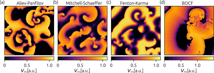
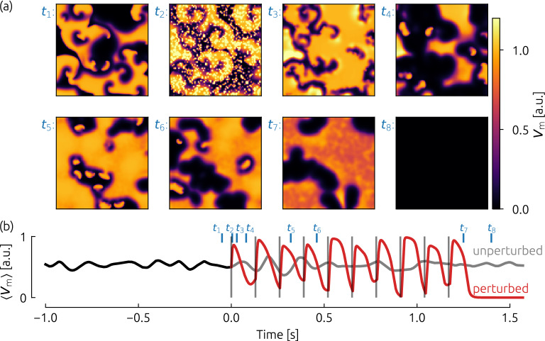
The conventional termination technique of life threatening cardiac arrhythmia like ventricular fibrillation is the application of a high-energy electrical defibrillation shock, coming along with severe side-effects. In order to improve the current treatment reducing these side-effects, the application of pulse sequences of lower energy instead of a single high-energy pulse are promising candidates. In this study, we show that in numerical simulations the dose-response function of pulse sequences applied to two-dimensional spiral wave chaos is not necessarily monotonously increasing, but exhibits a non-trivial frequency dependence. This insight into crucial phenomena appearing during termination attempts provides a deeper understanding of the governing termination mechanisms in general, and therefore may open up the path towards an efficient termination of cardiac arrhythmia in the future.


Conflict of interest statement

The authors declare no competing interests.

Figures

Figure 1
Figure 1
Exemplary snapshots of the membrane voltage Vm during episodes of spiral wave chaos are shown for the investigated cell models: the Aliev-Panfilov model AP (a), the Mitchell–Schaeffer model MS (b), the three-variable Fenton-Karma model FK (c), and the Bueno-Orovio-Cherry-Fenton model BOCF (d), respectively.
Figure 2
Figure 2
Exemplary (successful) termination attempt based on the Fenton-Karma model (FK) by a sequence of ten pulses. The frequency of the pulses in this example is equal to the dominant frequency fdom, and the amplitude of the pulses is E=0.5 a.u. In (a), eight snapshots of the membrane potential are shown just before the pacing protocol starts (t1), during the pacing (t2 - t6) and after the final pulse (t7 and t8). The spatially averaged membrane potential Vm (as a simple estimate for a pseudo ECG) is depicted in subplot (b), where pulses are marked with gray vertical lines and time instances which refer to the snapshots in (a) are marked in blue. From t=0 s, the red curve shows the case where the pacing protocol was applied, whereas the gray curve depicts the unperturbed case.
Figure 3
Figure 3
Overview of success rates depending on the pacing frequency. Each column depicts results for each investigated cardiac cell model, whereas the rows denote the number of pulses delivered, respectively (1 pulse, five pulses, ten pulses, and twenty pulses). The pacing frequency fpacing is given in multiples of the dominant frequency fdom which was determined for each cell model individually. Note that the frequency dependence for a single pulse is technically redundant, due to the lack of subsequent pulses. However, the data is still shown in order to demonstrate the magnitude of statistical fluctuations regarding the determination of the success rate. Horizontal lines at specific pacing frequencies denote dose-response curves, which are shown explicitly in Fig. 4. Two specific combinations of frequency and energies E1, and E2 (marked by white crosses) are discussed in more detail later.
Figure 4
Figure 4
Dose-Response curves for selected pacing frequencies ((a) AP: 1.3fdom, (b) MS: 1.1fdom, (c) FK: 1.1fdom, (d) BOCF: 1.04fdom). The success rates of the selected pacing frequencies are marked as horizontal gray dashed lines in Fig. 3.
Figure 5
Figure 5
The temporal evolution of the number of phase singularities (NPS) as a measure for the number of spiral waves in the system during a ten pulse protocol. For the Fenton-Karma model (FK), two pulse amplitudes were selected (E1=0.3 shown in (a) and E2=1.0 shown in (b)) for a specific pacing frequency of fpacing=1.1fdom (marked in Fig. 3 as white crosses). For these cases, the number of phase singularities was tracked over time for 200 simulations. After averaging over all simulations, the mean NPS is depicted in blue, whereas the standard deviation is marked in light blue.
Figure 6
Figure 6
Determination of the dominant frequency fdom of the system. The time series of the membrane potential was sampled for each cell model (a representative snapshot is shown in (a) for the FK model). The red and gray circles indicate two exemplary locations, where a fraction of the time series is shown in subplot (b). Based on these time series, Fourier spectra were computed (shown in subplot (c)), which were averaged in the end (black curve in (c)). Note, that in practice, this procedure was performed not just for two locations, but for all pixels of the spatial domain. Also, the temporal length of time series which were used for the computation of the Fourier amplitude spectra, was 20s for all cell models.

Similar articles

Cited by

References

    1. Wong CX, et al. Epidemiology of sudden cardiac death: Global and regional perspectives. Heart Lung Circ. 2019;28:6–14. doi: 10.1016/j.hlc.2018.08.026. - DOI - PubMed
    1. Winfree AT. Electrical instability in cardiac muscle: Phase singularities and rotors. J. Theor. Biol. 1989;138:353–405. doi: 10.1016/S0022-5193(89)80200-0. - DOI - PubMed
    1. Davidenko JM, Pertsov AV, Salomonsz R, Baxter W, Jalife J. Stationary and drifting spiral waves of excitation in isolated cardiac muscle. Nature. 1992;355:349–351. doi: 10.1038/355349a0. - DOI - PubMed
    1. Gray RA, Pertsov AM, Jalife J. Spatial and temporal organization during cardiac fibrillation. Nature. 1998;392:75–78. doi: 10.1038/32164. - DOI - PubMed
    1. Christoph J, et al. Electromechanical vortex filaments during cardiac fibrillation. Nature. 2018;555:667. doi: 10.1038/nature26001. - DOI - PubMed

Publication types