. 2022 Sep;47(10):1755-1763.
doi: 10.1038/s41386-022-01376-4. Epub 2022 Jul 14.

Prenatal opioid exposure inhibits microglial sculpting of the dopamine system selectively in adolescent male offspring

Affiliations

Prenatal opioid exposure inhibits microglial sculpting of the dopamine system selectively in adolescent male offspring

Caroline J Smith et al. Neuropsychopharmacology. 2022 Sep.

Abstract

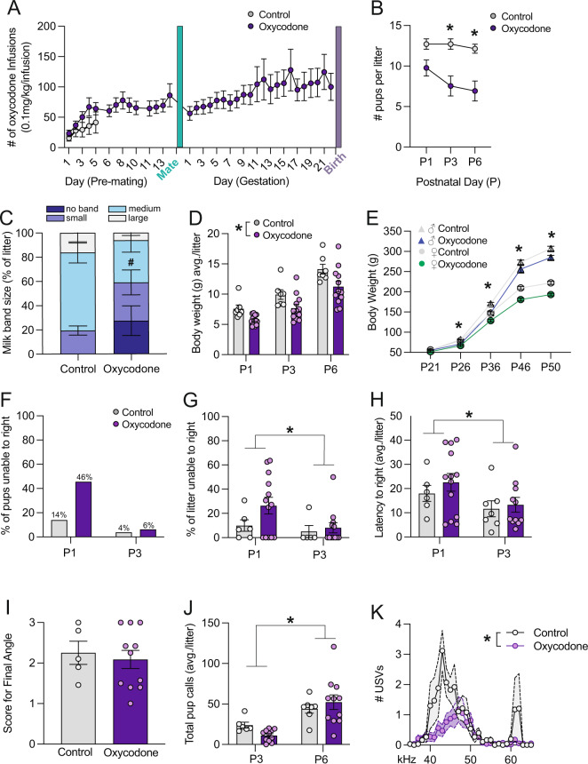
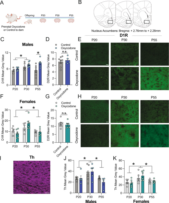
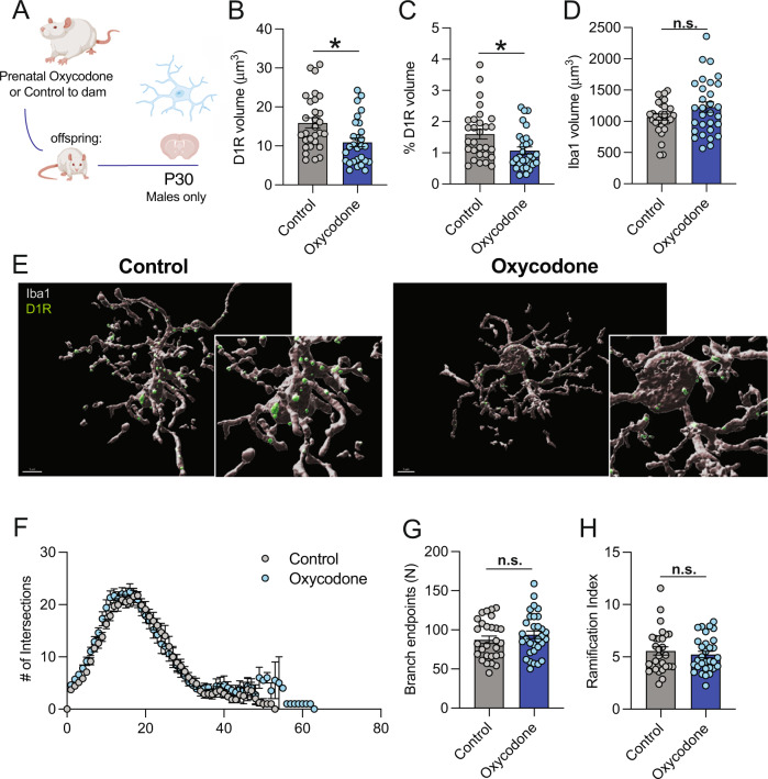
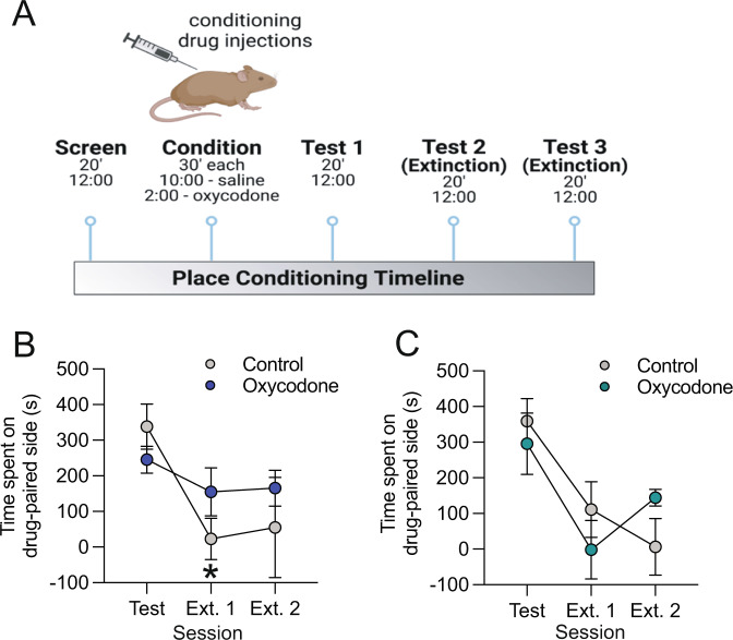
The current opioid epidemic has dramatically increased the number of children who are prenatally exposed to opioids, including oxycodone. A number of social and cognitive abnormalities have been documented in these children as they reach young adulthood. However, little is known about the mechanisms underlying developmental effects of prenatal opioid exposure. Microglia, the resident immune cells of the brain, respond to acute opioid exposure in adulthood. Moreover, microglia are known to sculpt neural circuits during typical development. Indeed, we recently found that microglial phagocytosis of dopamine D1 receptors (D1R) in the nucleus accumbens (NAc) is required for the natural developmental decline in NAc-D1R that occurs between adolescence and adulthood in rats. This microglial pruning occurs only in males, and is required for the normal developmental trajectory of social play behavior. However, virtually nothing is known as to whether this developmental program is altered by prenatal exposure to opioids. Here, we show in rats that maternal oxycodone self-administration during pregnancy leads to reduced adolescent microglial phagocytosis of D1R and subsequently higher D1R density within the NAc in adult male, but not female, offspring. Finally, we show prenatal and adult behavioral deficits in opioid-exposed offspring, including impaired extinction of oxycodone-conditioned place preference in males. This work demonstrates for the first time that microglia play a key role in translating prenatal opioid exposure to changes in neural systems and behavior.


Conflict of interest statement

The authors declare no competing interests.

Figures

Fig. 1
Fig. 1. Prenatal opioid exposure impacts neonatal outcomes in offspring.
A Rat dams self-administered oxycodone for either 5 days pre-pregnancy (control) or throughout gestation. B Number of pups per litter was significantly decreased at P3 and P6 following oxycodone exposure. C ~ 20% of oxyocodone exposed pups lacked a detectable milk band and oxycodone pups tended to have fewer medium-size milk bands. Data are presented as % in each rating category per litter. D We observed a main effect of treatment such that the average body weight of oxycodone exposed litters was lower than that of control litters. E Both male and female oxycodone-exposed offspring were persistently lower in body weight into adulthood. AD N = 7 litters (CON), 12 litters (Oxycodone), male and female offspring are combined. F 46% of oxycodone exposed pups were unable to right themselves in 60 s on P1, as compared to only 14% of control pups, N = 79–127 pups per group. G There was no significant treatment effect on the % of each litter unable to right or on the latency to right (H), N (litters) = 6 (P1 control), 5 (P3 control), 13 (P1 oxycodone), 12 (P3 oxycodone). However, both measures were significantly lower at P3 as compared to P1. I There was a significant main effect of age, but no significant main effect of treatment on total USVs at P3 and P6 (J). Analysis of USVs by frequency at P3 revealed significantly more USVs in control pups as compared to oxycodone-exposed pups, particularly at lower frequencies (K). Data represent Mean +/− SEM, *p = 0.05.
Fig. 2
Fig. 2. Prenatal oxycodone exposure increases D1R density in the NAc in adulthood in males but not females.
A Schematic of timeline for treatment and tissue collection. B Rat brain atlas images delineating NAc region where D1R, D2R, and Th were quantified (based on Paxinos and Watson Rat Brain Atlas). C In males, D1R mean grey value differed significantly with age, and was higher at P55 in oxycodone-exposed males as compared to control. D D2R did not differ with treatment at P55 in males. E Representative 20x images of D1R staining in the NAc of males, scale bar = 27 microns. F D1R peaked at P30 in both control and oxycodone-exposed females, but did not differ with treatment. G D2R did not differ with treatment at P55 in females. H Representative 20x images of D1R staining in the NAc of females, scale bar = 27 microns. I Representative 20x image of Th staining in the NAc of males, scale bar = 27 microns. J Th peaked at P30 in both control and oxycodone exposed males and females (K), but did not differ with treatment. Data represent Mean +/− SEM, *p = 0.05.
Fig. 3
Fig. 3. Prenatal opioid exposure decreases microglial engulfment of NAc-D1R during adolescence in males.
A Schematic of timeline for exposure and P30 engulfment assessment. B D1R volume within microglia was significantly decreased following oxycodone exposure in male offspring at P30. C % D1R volume of total microglial volume was significantly decreased following oxycodone exposure in male offspring at P30. D There was no treatment effect on microglial volume. E Representative 60x images of microglial D1R engulment: grey: Iba1, green: D1R, scale bar = 5 microns. F Scholl analysis revealed no differences in # of intersections between control and oxycodone males, or in branch endpoints (G) or Ramification Index (H). BH Dots represent individual microglia, N = 29 (control), 30 (oxycodone) nested per animal 6 and 7, respectively for analysis (nested t-tests). Data represent Mean +/− SEM, *p = 0.05.
Fig. 4
Fig. 4. Prenatal opioid exposure prevents extinction of oxycodone-conditioned place preference in adult male, but not female, offspring.
A Schematic of conditioned place preference procedure. B In males, a significant interaction effect was observed between treatment and session. Posthoc testing revealed significant group differences such that control male offspring showed a significantly lower preference for the drug-paired compartment on extinction session 1 as compared to the test session, while no decrease between test and extinction 1 was observed in oxycodone-exposed males. C Both control and oxycodone-exposed females showed extinction of conditioned place preference. Data represent Mean +/− SEM, *p = 0.05 relative to Test session.

References

    1. Haight SC, Ko JY, Tong VT, Bohm MK, Callaghan WM. Opioid use disorder documented at delivery hospitalization—United States, 1999–2014. MMWR Morb Mortal Wkly Rep. 2018;67:845–9. doi: 10.15585/mmwr.mm6731a1. - DOI - PMC - PubMed
    1. Conradt E, Flannery T, Aschner JL, Annett RD, Croen LA, Duarte CS.et al. Prenatal opioid exposure: neurodevelopmental consequences and future research priorities. Pediatrics. 2019;144: 10.1542/peds.2019-0128. - PMC - PubMed
    1. Arter S, Lambert J, Brokman A, Fall N. Diagnoses during the first three years of life for children with prenatal opioid exposure and neonatal abstinence syndrome using a large maternal-infant data hub. J Pediatr Nurs. 2021;61:34–9. doi: 10.1016/j.pedn.2021.03.011. - DOI - PubMed
    1. Nygaard E, Moe V, Slinning K, Walhovd KB. Longitudinal cognitive development of children born to mothers with opioid and polysubstance use. Pediatr Res. 2015;78:330–5. doi: 10.1038/pr.2015.95. - DOI - PMC - PubMed
    1. van Baar A, de Graaff BM. Cognitive development at preschool-age of infants of drug-dependent mothers. Dev Med Child Neurol. 1994;36:1063–75. doi: 10.1111/j.1469-8749.1994.tb11809.x. - DOI - PubMed

Publication types