

High D-glucose levels induce ACE2 expression via GLUT1 in human airway epithelial cell line Calu-3
- PMID: 35836103
- PMCID: PMC9282902
- DOI: 10.1186/s12860-022-00427-4
High D-glucose levels induce ACE2 expression via GLUT1 in human airway epithelial cell line Calu-3
Abstract
Background: Severe acute respiratory syndrome coronavirus 2 (SARS-CoV-2) enters the host cell by binding to angiotensin-converting enzyme 2 (ACE2) receptors. ACE2 is expressed on human airway epithelial cells. Increased ACE2 expression may be associated with potentially high risk of COVID-19. However, the factors responsible for the regulation of ACE2 expression in human airway epithelial cells are unknown. Furthermore, hyperglycemia is a risk factor for poor disease prognosis.
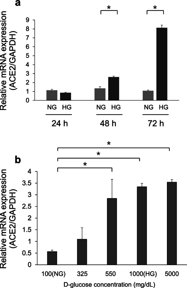
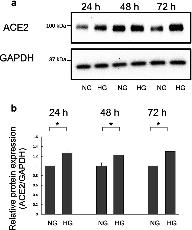
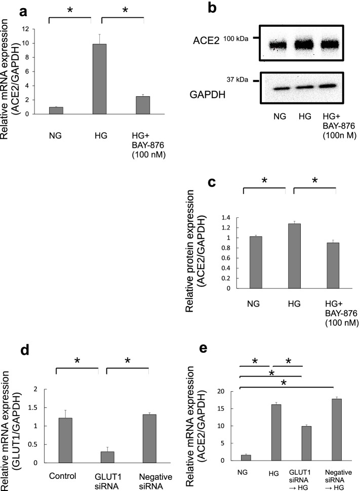
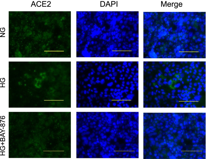
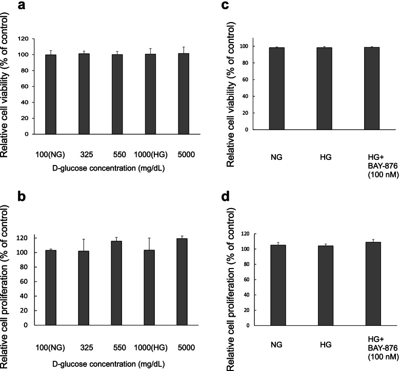
Results: In this study, we investigated the effects of D-glucose on ACE2 mRNA and protein expressions in Calu-3 bronchial submucosal cells. The cells were cultured in minimal essential medium containing different D-glucose concentrations. After 48 and 72 h of high D-glucose (1000 mg/dL) treatment, ACE2 mRNA expressions were significantly increased. ACE2 protein expressions were significantly increased after 24 h of high D-glucose treatment. ACE2 mRNA expression was enhanced by a D-glucose concentration of 550 mg/dL or more after 72 h of treatment. In addition, we investigated the role of glucose transporters (GLUTs) in Calu-3 cells. ACE2 mRNA and protein expressions were suppressed by the GLUT1 inhibitor BAY-876 in high D-glucose-treated Calu-3 cells. GLUT-1 siRNA was also used and ACE2 mRNA expressions were suppressed in high D-glucose-treated Calu-3 cells with GLUT-1 knockdown.
Conclusions: This is the first report indicating that high D-glucose levels induced ACE2 expression via GLUT1 in bronchial submucosal cells in vitro. As hyperglycemia can be treated appropriately, these findings could help reduce the risk of worsening of coronavirus disease 2019.
Keywords: ACE2; Angiotensin converting enzyme 2; COVID-19; Glucose transporters; SARS-CoV-2.
© 2022. The Author(s).
Conflict of interest statement
The authors declare that they have no competing interests.
Figures
Similar articles
-
Exercise-induced myokines downregulates the ACE2 level in bronchial epithelial cells: Implications for SARS-CoV-2 prevention.PLoS One. 2022 Jul 20;17(7):e0271303. doi: 10.1371/journal.pone.0271303. eCollection 2022. PLoS One. 2022. PMID: 35857747 Free PMC article.
-
Hypercapnia increases ACE2 expression and pseudo-SARS-CoV-2 entry in bronchial epithelial cells by augmenting cellular cholesterol.Front Immunol. 2023 Oct 12;14:1251120. doi: 10.3389/fimmu.2023.1251120. eCollection 2023. Front Immunol. 2023. PMID: 37901225 Free PMC article.
-
Effects of vitamin C and D on the mRNA expression of angiotensin converting enzyme 2 receptor, cathepsin L, and transmembrane serine protease in the mouse lungs.Libyan J Med. 2022 Dec;17(1):2054111. doi: 10.1080/19932820.2022.2054111. Libyan J Med. 2022. PMID: 35311495 Free PMC article.
-
Hyperglycemia, hydroxychloroquine, and the COVID-19 pandemic.J Med Virol. 2020 Jul;92(7):770-775. doi: 10.1002/jmv.25887. Epub 2020 Apr 27. J Med Virol. 2020. PMID: 32293710 Free PMC article. Review.
-
The pivotal link between ACE2 deficiency and SARS-CoV-2 infection.Eur J Intern Med. 2020 Jun;76:14-20. doi: 10.1016/j.ejim.2020.04.037. Epub 2020 Apr 20. Eur J Intern Med. 2020. PMID: 32336612 Free PMC article. Review.
Cited by
-
Getting sweeter: new evidence for glucose transporters in specific cell types of the airway?Am J Physiol Cell Physiol. 2023 Jan 1;324(1):C153-C166. doi: 10.1152/ajpcell.00140.2022. Epub 2022 Nov 21. Am J Physiol Cell Physiol. 2023. PMID: 36409177 Free PMC article. Review.
-
The Intersection of SARS-CoV-2 and Diabetes.Microorganisms. 2025 Jun 14;13(6):1390. doi: 10.3390/microorganisms13061390. Microorganisms. 2025. PMID: 40572277 Free PMC article. Review.
-
Molecular dissection of the role of ACE2 in glucose homeostasis.Physiol Rev. 2025 Jul 1;105(3):935-973. doi: 10.1152/physrev.00027.2024. Epub 2025 Feb 7. Physiol Rev. 2025. PMID: 39918873 Free PMC article. Review.
-
Anti-PD-L1 therapy altered inflammation but not survival in a lethal murine hepatitis virus-1 pneumonia model.Front Immunol. 2024 Jan 8;14:1308358. doi: 10.3389/fimmu.2023.1308358. eCollection 2023. Front Immunol. 2024. PMID: 38259435 Free PMC article.
-
Interactions of SARS-CoV-2 with Human Target Cells-A Metabolic View.Int J Mol Sci. 2024 Sep 16;25(18):9977. doi: 10.3390/ijms25189977. Int J Mol Sci. 2024. PMID: 39337465 Free PMC article. Review.
References
MeSH terms
Substances
LinkOut - more resources
Full Text Sources
Medical
Research Materials
Miscellaneous
