

Branch retinal artery occlusion caught in the act by an optical coherence tomography angiography image: case report
- PMID: 35836145
- PMCID: PMC9284791
- DOI: 10.1186/s12886-022-02517-5
Branch retinal artery occlusion caught in the act by an optical coherence tomography angiography image: case report
Abstract
Background: Retinal artery occlusion is a vascular entity caused by the temporary blockage of retinal arterioles.
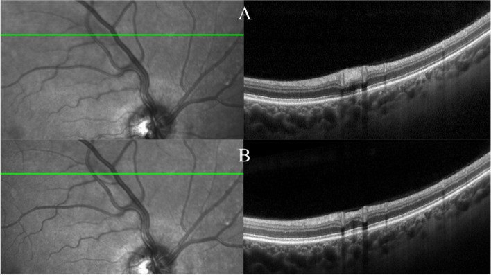
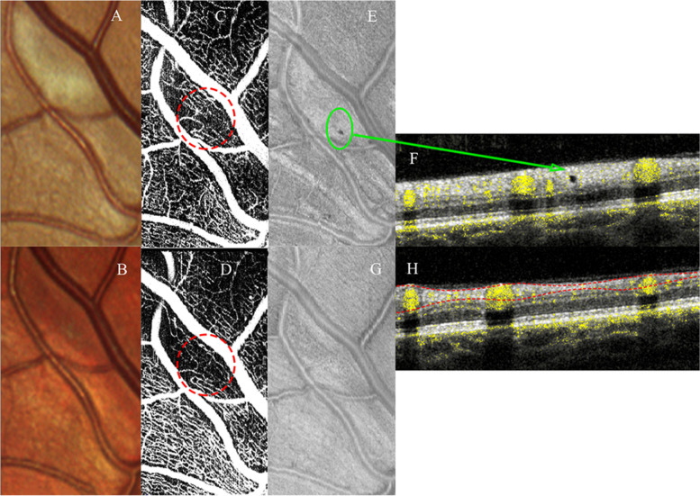
Case presentation: We present the case of a 57-year-old woman a partial visual loss in the right eye due to a cilioretinal artery occlusion. Ophthalmoscopy revealed a focal area of retinal whitening superior to the optic nerve in the right eye, while the left eye was within the limit. Retinal imaging, in particular optical coherence tomography angiography (OCTA), showed a capillary drop out of the superficial capillary plexus and the corresponding b-scan showed a round hyporeflective grey dot (optical empty) corresponding to the dark grey spot on the enface view at the level of the retinal whitening area.
Conclusion: Although the images did not allow the differentiation between vasospasm or retinal emboli, the OCTA imaging might help to identify and to caught in the act the specific region causing the retinal impairment. Also, the possible formation of small microcavity should be considered in case with branch retinal artery occlusion. The use of this new imaging technology might help to evaluate the efficacy of the therapy in vivo.
Keywords: Case report; Optical coherence tomography angiography (OCTA); Retinal artery occlusion; Spectral domain optical coherence tomography (SD-OCT).
© 2022. The Author(s).
Conflict of interest statement
Scarinci, Cacciamani and Ripandelli report no proprietary or financial interest to disclose.
Parravano reports personal fees from Allergan, from Bayer, and from Novartis outside the submitted work. The remaining authors declare that they have no competing interests.
Figures
Similar articles
-
CENTRAL RETINAL ARTERY OCCLUSION WITH DOUBLE CILIORETINAL ARTERY SPARING.Retin Cases Brief Rep. 2019 Winter;13(1):75-78. doi: 10.1097/ICB.0000000000000537. Retin Cases Brief Rep. 2019. PMID: 28085758
-
OPTICAL COHERENCE TOMOGRAPHY ANGIOGRAPHY SHOWS DEEP CAPILLARY PLEXUS HYPOPERFUSION IN INCOMPLETE CENTRAL RETINAL ARTERY OCCLUSION.Retin Cases Brief Rep. 2015 Fall;9(4):333-8. doi: 10.1097/ICB.0000000000000211. Retin Cases Brief Rep. 2015. PMID: 26355822
-
Incidental branch retinal artery occlusion on optical coherence tomography angiography presenting as segmental optic atrophy in a child: a case report.BMC Ophthalmol. 2017 Dec 19;17(1):256. doi: 10.1186/s12886-017-0653-6. BMC Ophthalmol. 2017. PMID: 29258533 Free PMC article.
-
Retinal vascular occlusion in pregnancy: three case reports and a review of the literature.J Med Case Rep. 2022 Apr 21;16(1):167. doi: 10.1186/s13256-022-03369-9. J Med Case Rep. 2022. PMID: 35449024 Free PMC article. Review.
-
Optical Coherence Tomography Angiography of Retinal Artery Occlusion.Dev Ophthalmol. 2016;56:122-31. doi: 10.1159/000442803. Epub 2016 Mar 15. Dev Ophthalmol. 2016. PMID: 27023788 Review.
References
Publication types
MeSH terms
LinkOut - more resources
Full Text Sources
