Review
. 2022 Dec;74(12):1169-1179.
doi: 10.1002/iub.2661. Epub 2022 Jul 14.

Cholesterol-dependent cytolysins: The outstanding questions

Affiliations
Review

Cholesterol-dependent cytolysins: The outstanding questions

Bronte A Johnstone et al. IUBMB Life. 2022 Dec.

Abstract

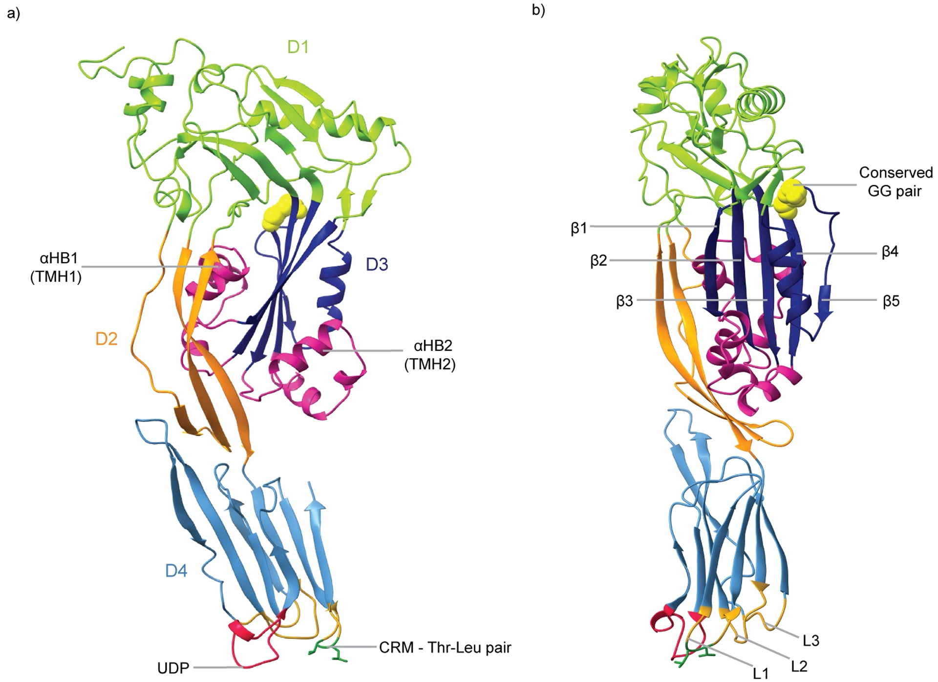
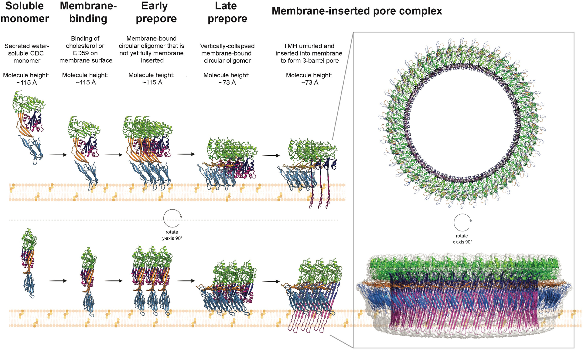
The cholesterol-dependent cytolysins (CDCs) are a major family of bacterial pore-forming proteins secreted as virulence factors by Gram-positive bacterial species. CDCs are produced as soluble, monomeric proteins that bind specifically to cholesterol-rich membranes, where they oligomerize into ring-shaped pores of more than 30 monomers. Understanding the details of the steps the toxin undergoes in converting from monomer to a membrane-spanning pore is a continuing challenge. In this review we summarize what we know about CDCs and highlight the remaining outstanding questions that require answers to obtain a complete picture of how these toxins kill cells.

Keywords: CD59; MACPF; cholesterol-binding protein; cholesterol-dependent cytolysin; intermedilysin; membrane-protein interactions; perfringolysin O; pneumolysin; pore-forming toxin.


Figures

Fig. 1
Fig. 1. Crystal structure of the CDC monomer
a) PFO monomeric structure (PDB ID: 1PFO) in which domains D1, D2, D3 and D4 are indicated in green, orange, navy and blue, respectively. The transmembrane hairpin (TMH) regions (αHB1 and αHB2) are colored in pink, the undecapeptide (UDP) in red, and the cholesterol recognition motif (CRM) in dark green. b) Side view of PFO (rotated by 90° about the y axis). The core beta-strands of D3 (β1 – β5) are indicated. The conserved glycine pair of D3 is shown in yellow CPK, and the loops L1 – L3 are in gold.
Fig. 2
Fig. 2. The mechanism for pore formation by CDCs
CDCs are produced and secreted as water-soluble, monomers. They bind cholesterol within cell membranes, or the receptor CD59 for some CDCs, triggering oligomerization into a membrane-tethered circular structure known as the prepore. Consisting of typically 35 – 45 monomers, this prepore stage can be divided into two separate events – the early prepore, characterized by a height of ~115 Å, and the subsequent late prepore at the lower height of ~73 Å, formed following a vertical collapse in height owing to rotation in D2. The final stage of the mechanism, membrane insertion, consists of the two α-helical bundles in each individual monomer undergoing a α-helix-to-β-strand transition and inserting into the membrane, forming the transmembrane β-hairpins of the β-barrel pore (> 250 Å in diameter). The figure was produced using the crystal structure of monomeric PLY (PDB ID: 5AOD) (69) and the fitted model of PLY for the cryo-EM structure of the PLY pore (PDB ID: 5LY6) (45).
Fig. 3
Fig. 3. Key interactions in prepore to pore conversion
Assembly of the prepore results in several critical interactions that stabilize the monomer-monomer interface and help drive the conformational changes that lead to pore formation. On opposite sides of the PFO molecule residues Y181, E183, F318 and K336 are brought together on adjacent monomers by oligomerization, resulting in a pair of intermolecular interactions – a salt bridge and an aromatic ring stack. The left-hand image shows PFO (PDB ID: 1PFO) as a cartoon colored by domain, with the four residues of interest shown as sticks, with the lower image a close-up on the D3 region of PFO to highlight the residues’ position. The right-hand image is of a pair of PFO monomers from a model of the PFO pore, built on the basis of the cryo-EM structure of the PLY pore (45). The inset again shows a close-up view of the residues, but in this case in the interface between the two monomers. The network of interactions between the residues in this interface is clearly visible.

References

    1. Hotze EM. and Tweten RK (2012) Membrane assembly of the cholesterol-dependent cytolysin pore complex. Biochim. Biophys. Acta, Biomembr 1818, 1028–1038. - PMC - PubMed
    1. Hotze EM, Le HM, Sieber JR, Bruxvoort C, McInerney MJ, et al. (2013) Identification and characterization of the first cholesterol-dependent cytolysins from Gram-negative bacteria. Infect. Immun 81, 216–225. - PMC - PubMed
    1. Gelber SE, Aguilar JL, Lewis KL, and Ratner AJ (2008) Functional and phylogenetic characterization of vaginolysin, the human-specific cytolysin from Gardnerella vaginalis. J. Bacteriol 190, 3896–3903. - PMC - PubMed
    1. Giddings KS, Zhao J, Sims PJ, and Tweten RK (2004) Human CD59 is a receptor for the cholesterol-dependent cytolysin intermedilysin. Nat. Struct. Mol. Biol 11, 1173–1178. - PubMed
    1. Wickham SE, Hotze EM, Farrand AJ, Polekhina G, Nero TL, et al. (2011) Mapping the intermedilysin-human CD59 receptor interface reveals a deep correspondence with the binding site on CD59 for complement binding proteins C8alpha and C9. J. Biol. Chem 286, 20952–20962. - PMC - PubMed

Publication types

MeSH terms

LinkOut - more resources