. 2022 Jun 8;24(2):498.
doi: 10.3892/etm.2022.11425. eCollection 2022 Aug.

A novel nude mouse model for studying the pathogenesis of endometriosis

Affiliations

A novel nude mouse model for studying the pathogenesis of endometriosis

Li-Ping Li et al. Exp Ther Med. .

Abstract

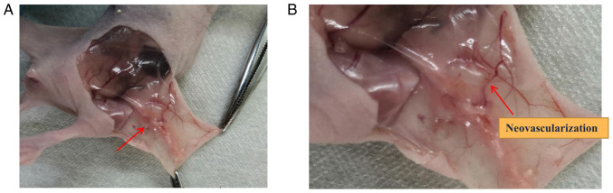
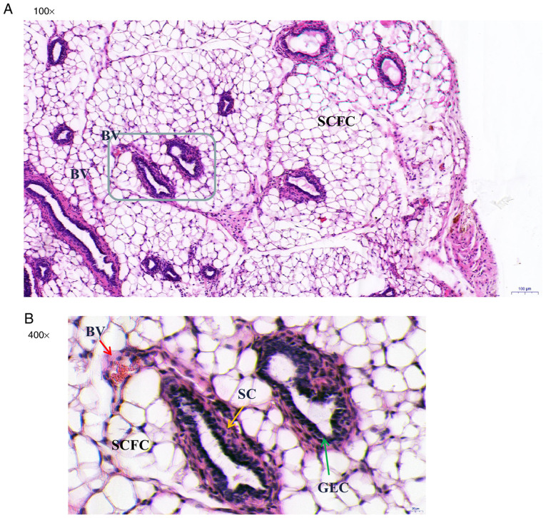
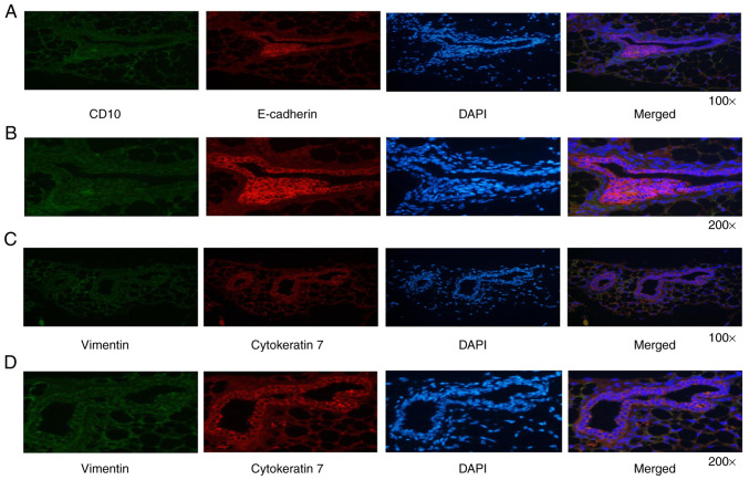
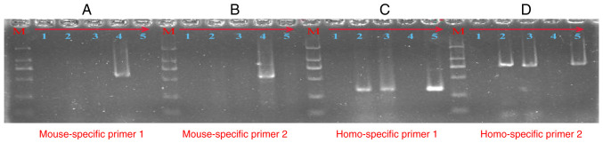
Endometriosis is a common female gynecological disease that is characterized by the presence of functional endometrial tissue outside the uterine cavity. At present, many animal models have been established. However, previous studies consistently use human endometrial tissue implanted in the subcutaneous or abdominal cavity for modeling and rarely use endometrial cells. In the present study, we ascertained whether immortalized stromal and/or epithelial endometrial cells are able to induce subcutaneous endometriosis in nude mice. Mixed human immortalized endometriosis stromal and epithelial cells, but not the cells of Group 1 or Group 2, were successfully constructed and led to endometriotic-like lesions. The endometriosis-like lesions observed in nude mice consisted of endometriosis-like glands lined with columnar epithelial cells and surrounded by stromal cells in the fibrous fatty connective tissue. Immunofluorescence analysis showed that glandular epithelial cells were intensely stained for E-cadherin and cytokeratin 7, and surrounding stromal cells were mildly stained for neprilysin (CD10) and vimentin. Moreover, the cells present in the endometriosis-like lesions were of human origin. Our data indicate that the mixture of human immortalized endometriosis stromal cells and epithelial cells is able to establish subcutaneous endometriosis lesions in nude mice. This model could be used to understand the molecular mechanisms involved in the occurrence and development of endometriosis.

Keywords: T HESCs; endometriosis; epithelial cells; nude mice; subcutaneous endometriotic-like lesions.


Conflict of interest statement

The authors declare that they have no competing interests.

Figures

Figure 1
Figure 1
Representative photo images of endometriosis-like lesions in Group 3. (А) Original image; arrow indicates the endometrioid lesion. (В) Partial enlarged image; arrow indicates the blood vessel formation. Group 3: 2x10⁶ T HESCs+2x10⁶ EECs.
Figure 2
Figure 2
Histologic characteristics of endometriosis-like lesions in nude mice, detected by hematoxylin and eosin (H&E) staining. Cross section of embedded endometriosis-like lesions is shown at original magnifications of x100 and x400. (A) Note the presence of endometriosis glands (grey-lined area) in the fibrous fatty connective tissues. (B) Developed and organized endometriosis glands with acini lined with GEC (green arrow). These endometriosis-like glands are lined with columnar epithelial cells and surrounded by SC (yellow arrow). The BV (red arrow) are filled with red blood cells and lined with flattened endothelial cells. BV, blood vessels; SCFC, subcutaneous fat cells; GEC, glandular epithelial cells; SC, stromal cells.
Figure 3
Figure 3
Immunofluorescence characterization of the endometriosis-like lesions. (A and B) Glandular epithelial cells were intensely stained for E-cadherin and surrounding stromal cells were mildly stained for CD10. (C and D) Glandular epithelial cells were intensely stained for cytokeratin 7 and surrounding stromal cells were mildly stained for vimentin.
Figure 4
Figure 4
Agarose gel electrophoresis analysis for the PCR products. The 571-bp (A) and 549-bp (B) PCR products were amplified by mouse-specific GAPDH primers. The 327-bp (C) and 772-bp (D) PCR products were amplified by human-specific GAPDH primers. Lanes 1-5, respectively, represent: the no-template control, DNA from endometriosis-like lesion tissue (no. 2), DNA from endometriosis-like lesion tissue (no. 3), positive control of mouse DNA, positive control of human peripheral blood DNA. Lane M is the DNA DL2000 marker: 2000 bp\1000 bp\750 bp\500 bp\250 bp\100 bp. GAPDH, glyceraldehyde 3-phosphate dehydrogenase.

Similar articles

Cited by

References

    1. Koninckx PR, Ussia A, Adamyan L, Wattiez A, Gomel V, Martin DC. Pathogenesis of endometriosis: The genetic/epigenetic theory. Fertil Steril. 2019;111:327–340. doi: 10.1016/j.fertnstert.2018.10.013. - DOI - PubMed
    1. Rolla E. Endometriosis: Advances and controversies in classification, pathogenesis, diagnosis, and treatment. F1000Res 8: F1000 Faculty Rev-529, 2019. - PMC - PubMed
    1. Zondervan KT, Becker CM, Missmer SA. Endometriosis. N Engl J Med. 2020;382:1244–1256. doi: 10.1056/NEJMra1810764. - DOI - PubMed
    1. Czyzyk A, Podfigurna A, Szeliga A, Meczekalski B. Update on endometriosis pathogenesis. Minerva Ginecol. 2017;69:447–461. doi: 10.23736/S0026-4784.17.04048-5. - DOI - PubMed
    1. Vercellini P, Vigano P, Somigliana E, Fedele L. Endometriosis: Pathogenesis and treatment. Nat Rev Endocrinol. 2014;10:261–275. doi: 10.1038/nrendo.2013.255. - DOI - PubMed