

Functional Regulation of KATP Channels and Mutant Insight Into Clinical Therapeutic Strategies in Cardiovascular Diseases
- PMID: 35837280
- PMCID: PMC9274113
- DOI: 10.3389/fphar.2022.868401
Functional Regulation of KATP Channels and Mutant Insight Into Clinical Therapeutic Strategies in Cardiovascular Diseases
Abstract
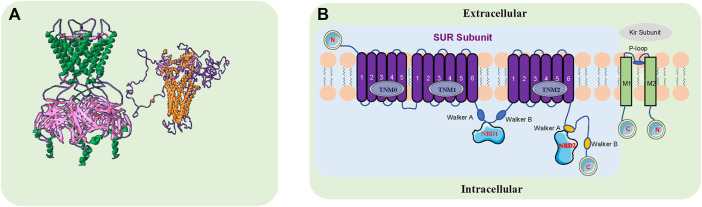
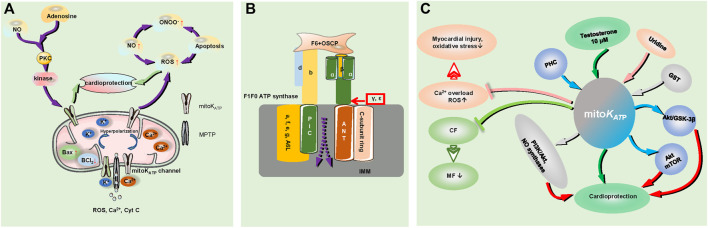
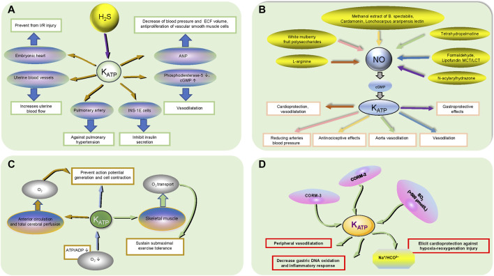
ATP-sensitive potassium channels (KATP channels) play pivotal roles in excitable cells and link cellular metabolism with membrane excitability. The action potential converts electricity into dynamics by ion channel-mediated ion exchange to generate systole, involved in every heartbeat. Activation of the KATP channel repolarizes the membrane potential and decreases early afterdepolarization (EAD)-mediated arrhythmias. KATP channels in cardiomyocytes have less function under physiological conditions but they open during severe and prolonged anoxia due to a reduced ATP/ADP ratio, lessening cellular excitability and thus preventing action potential generation and cell contraction. Small active molecules activate and enhance the opening of the KATP channel, which induces the repolarization of the membrane and decreases the occurrence of malignant arrhythmia. Accumulated evidence indicates that mutation of KATP channels deteriorates the regulatory roles in mutation-related diseases. However, patients with mutations in KATP channels still have no efficient treatment. Hence, in this study, we describe the role of KATP channels and subunits in angiocardiopathy, summarize the mutations of the KATP channels and the functional regulation of small active molecules in KATP channels, elucidate the potential mechanisms of mutant KATP channels and provide insight into clinical therapeutic strategies.
Keywords: KATP channels; channelopathy; mitoKATP channels; mutation; myocardial ischemia; small active molecules.
Copyright © 2022 Wang, Bian, Yan and Zhang.
Conflict of interest statement
The authors declare that the research was conducted in the absence of any commercial or financial relationships that could be construed as a potential conflict of interest.
Figures
Similar articles
-
Molecular biology of adenosine triphosphate-sensitive potassium channels.Endocr Rev. 1999 Apr;20(2):101-35. doi: 10.1210/edrv.20.2.0361. Endocr Rev. 1999. PMID: 10204114 Review.
-
Electrophysiological analysis of cardiac KATP channel.Biophys Rep. 2025 Apr 30;11(2):77-86. doi: 10.52601/bpr.2024.240023. Biophys Rep. 2025. PMID: 40308939 Free PMC article.
-
KATP channels and cardioprotection.Arh Farm (Belgr). 2024;74(5):625-657. doi: 10.5937/arhfarm74-51604. Epub 2024 Nov 1. Arh Farm (Belgr). 2024. PMID: 40400696
-
Metabolic regulation of cardiac ATP-sensitive K+ channels.Cardiovasc Drugs Ther. 1993 Aug;7 Suppl 3:499-505. doi: 10.1007/BF00877614. Cardiovasc Drugs Ther. 1993. PMID: 8251419
-
Potassium channel openers as potential therapeutic weapons in ion channel disease.Kidney Int. 2000 Mar;57(3):838-45. doi: 10.1046/j.1523-1755.2000.00923.x. Kidney Int. 2000. PMID: 10720937 Review.
Cited by
-
Motor Dysfunction of Gastric Antral Smooth Muscle in Diabetic Rats: Contribution of ATP-Dependent Potassium Channels.Adv Biomed Res. 2023 Jul 27;12:199. doi: 10.4103/abr.abr_44_23. eCollection 2023. Adv Biomed Res. 2023. PMID: 37694236 Free PMC article.
-
Nicorandil Exerts Anticonvulsant Effects in Pentylenetetrazol-Induced Seizures and Maximal-Electroshock-Induced Seizures by Downregulating Excitability in Hippocampal Pyramidal Neurons.Neurochem Res. 2023 Sep;48(9):2701-2713. doi: 10.1007/s11064-023-03932-w. Epub 2023 Apr 19. Neurochem Res. 2023. PMID: 37076745
-
miRNA Expression Profiles in Isolated Ventricular Cardiomyocytes: Insights into Doxorubicin-Induced Cardiotoxicity.Int J Mol Sci. 2024 May 12;25(10):5272. doi: 10.3390/ijms25105272. Int J Mol Sci. 2024. PMID: 38791311 Free PMC article.
References
-
- Akhtar M. M., Lorenzini M., Pavlou M., Ochoa J. P., O'Mahony C., Restrepo-Cordoba M. A., et al. (2021). Association of Left Ventricular Systolic Dysfunction Among Carriers of Truncating Variants in Filamin C with Frequent Ventricular Arrhythmia and End-Stage Heart Failure. JAMA Cardiol. 6 (8), 891–901. 10.1001/jamacardio.2021.1106 - DOI - PMC - PubMed
-
- Assreuy A. M. S., Amorim R. M. F., Martins S. L., de Queiroz Martins M. G., Cajazeiras J. B., da Silva M. T. L., et al. (2020). Antinociceptive Effect of Lonchocarpus Araripensis Lectin: Activation of L-arginine/NO/cGMP/K+ATP Signaling Pathway. Inflammopharmacology 28 (6), 1623–1631. 10.1007/s10787-020-00729-z - DOI - PubMed
-
- Balamurugan K., Kavitha B., Yang Z., Mohan V., Radha V., Shyng S. L. (2019). Functional Characterization of Activating Mutations in the Sulfonylurea Receptor 1 (ABCC8) Causing Neonatal Diabetes Mellitus in Asian Indian Children. Pediatr. Diabetes 20 (4), 397–407. 10.1111/pedi.12843 - DOI - PMC - PubMed
Publication types
LinkOut - more resources
Full Text Sources