. 2022 Jun 28:13:930041.
doi: 10.3389/fphar.2022.930041. eCollection 2022.

Comprehensive Analysis of Cuproptosis-Related Genes in Immune Infiltration and Prognosis in Melanoma

Affiliations

Comprehensive Analysis of Cuproptosis-Related Genes in Immune Infiltration and Prognosis in Melanoma

Haozhen Lv et al. Front Pharmacol. .

Abstract

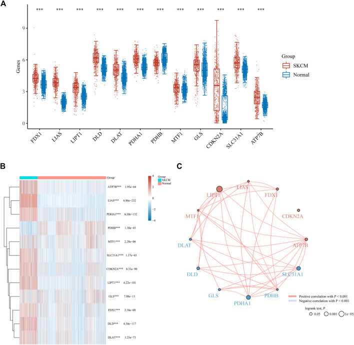
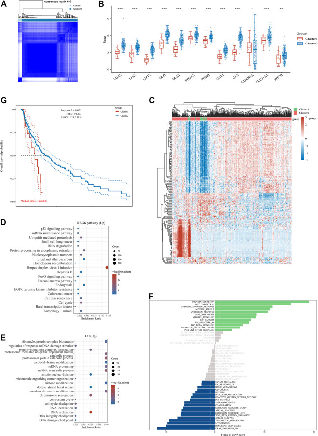
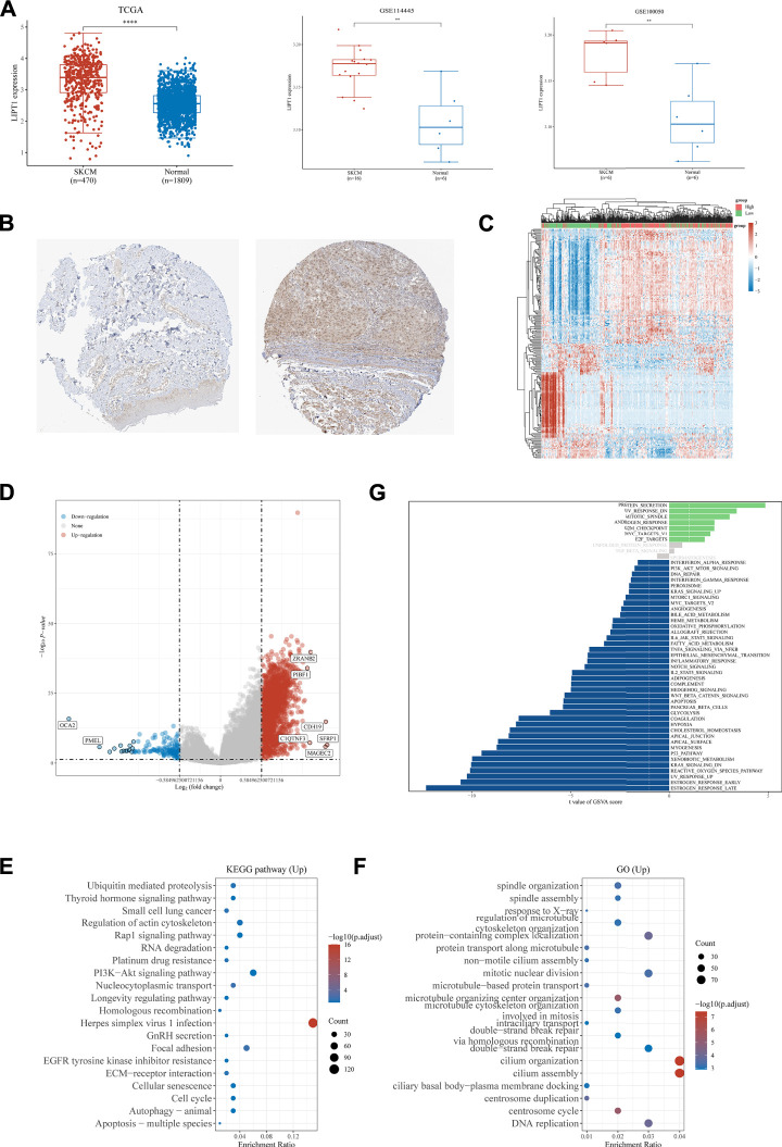
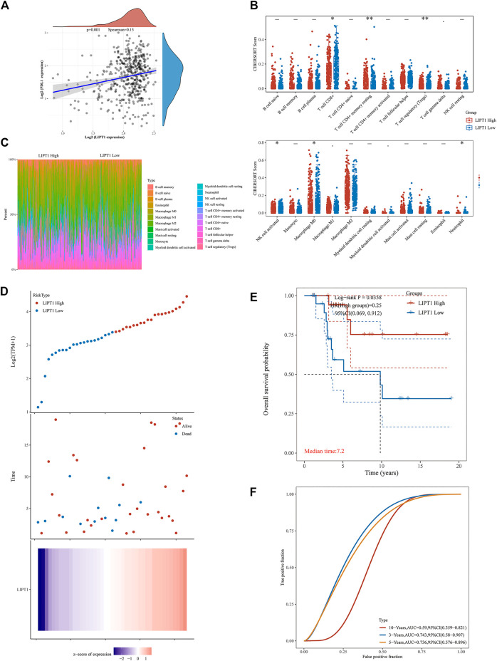
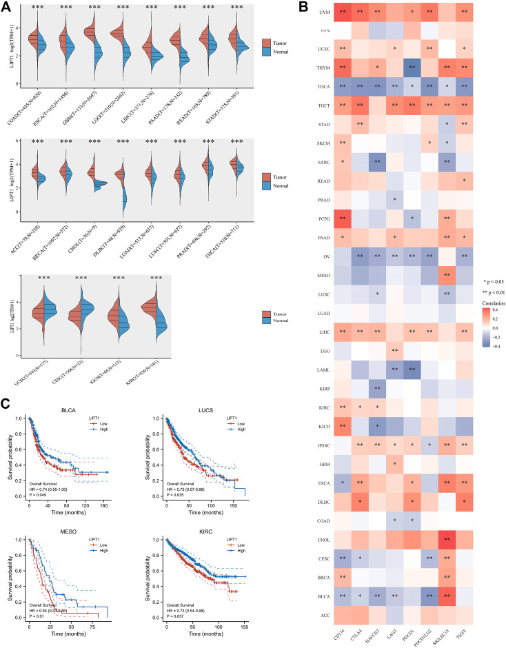
Skin cutaneous melanoma (SKCM, hereafter referred to as melanoma) is the most lethal skin cancer with increasing incidence. Regulated cell death plays an important role in tumorigenesis and serves as an important target for almost all treatment strategies. Cuproptosis is the most recently identified copper-dependent regulated cell death form that relies on mitochondria respiration. However, its role in tumorigenesis remains unknown. The correlation of cuproptosis-related genes with tumor prognosis is far to be understood, either. In the present study, we explored the correlation between cuproptosis-related genes with the prognosis of melanoma by accessing and analyzing a public database and found 11 out 12 genes were upregulated in melanoma tissues and three genes (LIPT1, PDHA1, and SLC31A1) have predictive value for the prognosis. The subgroup of melanoma patients with higher cuproptosis-related gene expression showed longer overall survival than those with lower gene expression. We chose LIPT1 for further exploration. LIPT1 expression was increased in melanoma biopsies and was an independent favorable prognostic indicator for melanoma patients. Moreover, LIPT1 expression was positively correlated with PD-L1 expression and negatively associated with Treg cell infiltration. The melanoma patients with higher LIPT1 expression showed longer overall survival than those with lower LIPT1 expression after receiving immunotherapy, indicating the prognostic predictive value of LIPT1. Finally, a pan-cancer analysis indicated that LIPT1 was differentially expressed in diverse cancers as compared to normal tissues and correlated with the expression of multiple immune checkpoints, especially PD-L1. It could serve as a favorable prognosis indicator in some cancer types. In conclusion, our study demonstrated the prognostic value of cuproptosis-related genes, especially LIPT1, in melanoma, and revealed the correlation between LIPT1 expression and immune infiltration in melanoma, thus providing new clues on the prognostic assessment of melanoma patients and providing a new target for the immunotherapy of melanoma.

Keywords: LIPT1; immune infiltration; ion-driven cell death; melanoma; prognosis.


Conflict of interest statement

The authors declare that the research was conducted in the absence of any commercial or financial relationships that could be construed as a potential conflict of interest.

Figures

FIGURE 1
FIGURE 1
Cuproptosis-related genes expressed in SKCM patients and their correlation and prognostic value. Box plots (A) and heat map (B) of cuproptosis-related genes in SKCM compared to normal tissues. (C) Spearman correlation and prognostic values of cuproptosis-related genes in SKCM. Red represents positive correlation whereas blue represents negative correlation. The larger the circle the smaller log-rank p. ***p < 0.001.
FIGURE 2
FIGURE 2
Differential expression pattern of cuproptosis-related genes and survival and functional enrichment analysis in two Skin Cutaneous Melanoma (SKCM) clusters. (A) Consensus clustering matrix for k = 2. (B) Box plots visualized the expression patterns of cuproptosis-related genes in two SKCM clusters. (C) The heatmap of the differential gene expression between two SKCM clusters. (D–F) The most significant KEGG pathways (D), GO enrichment (E) and activation of multiple pathways by GSVA (F) in two SKCM clusters. (G) The Kaplan–Meier curves show the overall survival for two clusters of SKCM patients. **p < 0.01, and ***p < 0.001.
FIGURE 3
FIGURE 3
Analysis of the LIPT1 gene expression in SKCM. (A) Compared to normal tissues, LIPT1 was upregulated in SKCM tissues in the TCGA database, GSE114445, and GSE100050. (B) Immunohistochemistry analysis showed that LIPT1 was highly expressed in SKCM tissue (right) compared with normal skin tissue (left) in the human protein atlas (antibody HPA034802). The heatmap (C) and Volcano plot (D) of the differential gene expression between high and low expressed LIPT1 in SKCM. (E,F) The most significant KEGG pathways (E) and GO enrichment (F) between high and low expressed LIPT1 in SKCM. (G) Up and downregulation of LIPT1 was associated with activation of multiple pathways by GSVA. **p < 0.01 and ****p < 0.0001.
FIGURE 4
FIGURE 4
Upregulated LIPT1 expression is connection with good outcomes of Skin Cutaneous Melanoma (SKCM). (A) The Kaplan–Meier analysis of SKCM patients with high and low LIPT1 expression level in the TCGA cohort. (B,C) Univariate (B) and multivariate Cox analysis of expression of LIPT1 and other factors in SKCM patients.
FIGURE 5
FIGURE 5
Relationship between LIPT1 expression, immunotherapy and infiltrating immune cells in Skin Cutaneous Melanoma (SKCM) by correlation analysis. (A) The correlation analysis of PD-L1 expression and LIPT1 expression in SKCM. (B) The infiltrating levels of immune cells in high and low LIPT1 expression groups in SKCM patients. (C) The percentage abundance of tumor-infiltrating immune cells showed the immune infiltration analysis between high and low LIPT1 expression groups in SKCM patients. (D) The gene expression, survival time, and survival status of the TCGA database. (E) Kaplan-Meier survival analysis of the gene signature from TCGA cohort. (F) The ROC curve of LIPT1.
FIGURE 6
FIGURE 6
Comprehensive analysis of LIPT1 in pan-cancers. (A) LIPT1 was differentially expressed in most cancer types. (C) Highly expressed LIPT1 showed good overall survival in multiple cancers. (B) The relationship between expression of LIPT1 and eight common immune checkpoints in pan-cancers. *p < 0.05, **p < 0.01, and ***p < 0.001.

References

    1. Arce Vargas F., Furness A. J. S., Solomon I., Joshi K., Mekkaoui L., Lesko M. H., et al. (2017). Fc-Optimized Anti-CD25 Depletes Tumor-Infiltrating Regulatory T Cells and Synergizes with PD-1 Blockade to Eradicate Established Tumors. Immunity 46 (4), 577–586. 10.1016/j.immuni.2017.03.013 - DOI - PMC - PubMed
    1. Ayers M., Lunceford J., Nebozhyn M., Murphy E., Loboda A., Kaufman D. R., et al. (2017). IFN-γ-related mRNA Profile Predicts Clinical Response to PD-1 Blockade. J. Clin. Invest. 127 (8), 2930–2940. 10.1172/JCI91190 - DOI - PMC - PubMed
    1. Bagchi S., Yuan R., Engleman E. G. (2021). Immune Checkpoint Inhibitors for the Treatment of Cancer: Clinical Impact and Mechanisms of Response and Resistance. Annu. Rev. Pathol. 16, 223–249. 10.1146/annurev-pathol-042020-042741 - DOI - PubMed
    1. Banach K., Kowalska J., Rzepka Z., Beberok A., Rok J., Wrześniok D. (2021). The Role of UVA Radiation in Ketoprofen-Mediated BRAF-Mutant Amelanotic Melanoma Cells Death - A Study at the Cellular and Molecular Level. Toxicol. In Vitro 72, 105108. 10.1016/j.tiv.2021.105108 - DOI - PubMed
    1. Barrios D. M., Do M. H., Phillips G. S., Postow M. A., Akaike T., Nghiem P., et al. (2020). Immune Checkpoint Inhibitors to Treat Cutaneous Malignancies. J. Am. Acad. Dermatol 83 (5), 1239–1253. 10.1016/j.jaad.2020.03.131 - DOI - PMC - PubMed