

Overexpression of LIMA1 Indicates Poor Prognosis and Promotes Epithelial-Mesenchymal Transition in Head and Neck Squamous Cell Carcinoma
- PMID: 35837368
- PMCID: PMC9274436
- DOI: 10.1177/11795549221109493
Overexpression of LIMA1 Indicates Poor Prognosis and Promotes Epithelial-Mesenchymal Transition in Head and Neck Squamous Cell Carcinoma
Abstract
Background: LIMA1 encodes LIM domain and actin binding 1, a cytoskeleton-associated protein whose loss has been linked to migration and invasion behavior of cancer cells. However, the roles of LIMA1 underlying the malignant behavior of tumors in head and neck squamous cell carcinoma (HNSC) are not fully understood.
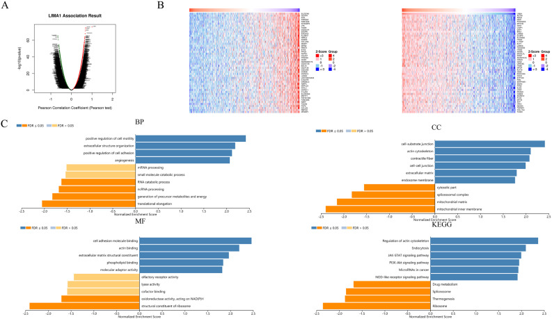
Methods: We conducted a multi-omics study on the role of LIMA1 in HNSC based on The Cancer Genome Atlas data. Subsequent in vitro experiments were performed to validate the results of bioinformatic analysis. We first identified the correlation between LIMA1 and tumor cell functional states according to single-cell sequencing data in HNSC. The potential downstream effects of LIMA1 were explored for gene ontology and Kyoto Encyclopedia of Genes and Genomes pathways through functional enrichment analysis of the gene sets that correlated with LIMA1 in HNSC. The prognostic role of LIMA1 was assessed using the log rank test to compare difference in survival between LIMA1High and LIMA1Low patients. Univariate Cox regression and multivariate Cox regression were further carried out to identify the prognostic value of LIMA1 in HNSC.
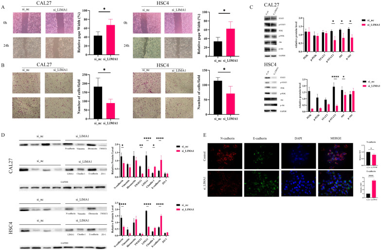
Results: LIMA1 was identified as a prognostic biomarker and is associated with epithelial-mesenchymal transition (EMT) progress in HNSC. In vitro silencing of LIMA1 suppressed EMT and related pathways in HNSC.
Conclusions: LIMA1 promotes EMT and further leads to tumor invasion and metastasis. Increased expression of LIMA1 indicates poor survival, identifying it as a prognostic biomarker in HNSC.
Keywords: LIMA1; TCGA; epithelial-mesenchymal transition; head and neck squamous cell carcinoma; prognosis.
© The Author(s) 2022.
Conflict of interest statement
Declaration of Conflicting Interests: The author(s) declared no potential conflicts of interest with respect to the research, authorship, and/or publication of this article.
Figures
Similar articles
-
Characterization of LIMA1 and its emerging roles and potential therapeutic prospects in cancers.Front Oncol. 2023 May 19;13:1115943. doi: 10.3389/fonc.2023.1115943. eCollection 2023. Front Oncol. 2023. PMID: 37274282 Free PMC article. Review.
-
The Relationship between the Prognostic Marker LIMA1 in Head and Neck Squamous Cell Carcinoma and Immune Infiltration.J Oncol. 2022 Aug 31;2022:1040116. doi: 10.1155/2022/1040116. eCollection 2022. J Oncol. 2022. PMID: 37181789 Free PMC article.
-
Prognostic value of CD247 in patients with head and neck squamous cell carcinoma: bioinformatic analysis of TCGA database.Ann Transl Med. 2022 Sep;10(17):923. doi: 10.21037/atm-22-1143. Ann Transl Med. 2022. PMID: 36172089 Free PMC article.
-
Identification of SLC2A3 as a prognostic indicator correlated with the NF-κB/EMT axis and immune response in head and neck squamous cell carcinoma.Channels (Austin). 2023 Dec;17(1):2208928. doi: 10.1080/19336950.2023.2208928. Channels (Austin). 2023. PMID: 37134043 Free PMC article.
-
Prognostic value of epithelial-mesenchymal transition-inducing transcription factors in head and neck squamous cell carcinoma: A meta-analysis.Head Neck. 2020 May;42(5):1067-1076. doi: 10.1002/hed.26104. Epub 2020 Feb 12. Head Neck. 2020. PMID: 32048783 Review.
Cited by
-
Exploring the Enigma: The Role of the Epithelial Protein Lost in Neoplasm in Normal Physiology and Cancer Pathogenesis.Int J Mol Sci. 2024 May 2;25(9):4970. doi: 10.3390/ijms25094970. Int J Mol Sci. 2024. PMID: 38732188 Free PMC article. Review.
-
M6A modification-mediated LIMA1 promotes the progression of hepatocellular carcinoma through the wnt-βcatenin/Hippo pathway.Cell Biol Toxicol. 2024 Dec 21;41(1):9. doi: 10.1007/s10565-024-09959-1. Cell Biol Toxicol. 2024. PMID: 39707043 Free PMC article.
-
Expression and molecular insights of lima1 in cholangiocarcinoma.Cell Adh Migr. 2024 Dec;18(1):4-17. doi: 10.1080/19336918.2024.2383068. Epub 2024 Jul 30. Cell Adh Migr. 2024. PMID: 39076043 Free PMC article.
-
LIMA1-alpha staining predicts curative intent surgery response in HPV negative head and neck cancer.EMBO Mol Med. 2025 Aug;17(8):2095-2114. doi: 10.1038/s44321-025-00266-8. Epub 2025 Jul 17. EMBO Mol Med. 2025. PMID: 40676267 Free PMC article.
-
Characterization of LIMA1 and its emerging roles and potential therapeutic prospects in cancers.Front Oncol. 2023 May 19;13:1115943. doi: 10.3389/fonc.2023.1115943. eCollection 2023. Front Oncol. 2023. PMID: 37274282 Free PMC article. Review.
References
LinkOut - more resources
Full Text Sources
