Review
. 2022 Jun 28:10:901317.
doi: 10.3389/fbioe.2022.901317. eCollection 2022.

Gene Therapy in Orthopaedics: Progress and Challenges in Pre-Clinical Development and Translation

Affiliations
Review

Gene Therapy in Orthopaedics: Progress and Challenges in Pre-Clinical Development and Translation

Rachael S Watson-Levings et al. Front Bioeng Biotechnol. .

Abstract

In orthopaedics, gene-based treatment approaches are being investigated for an array of common -yet medically challenging- pathologic conditions of the skeletal connective tissues and structures (bone, cartilage, ligament, tendon, joints, intervertebral discs etc.). As the skeletal system protects the vital organs and provides weight-bearing structural support, the various tissues are principally composed of dense extracellular matrix (ECM), often with minimal cellularity and vasculature. Due to their functional roles, composition, and distribution throughout the body the skeletal tissues are prone to traumatic injury, and/or structural failure from chronic inflammation and matrix degradation. Due to a mixture of environment and endogenous factors repair processes are often slow and fail to restore the native quality of the ECM and its function. In other cases, large-scale lesions from severe trauma or tumor surgery, exceed the body's healing and regenerative capacity. Although a wide range of exogenous gene products (proteins and RNAs) have the potential to enhance tissue repair/regeneration and inhibit degenerative disease their clinical use is hindered by the absence of practical methods for safe, effective delivery. Cumulatively, a large body of evidence demonstrates the capacity to transfer coding sequences for biologic agents to cells in the skeletal tissues to achieve prolonged delivery at functional levels to augment local repair or inhibit pathologic processes. With an eye toward clinical translation, we discuss the research progress in the primary injury and disease targets in orthopaedic gene therapy. Technical considerations important to the exploration and pre-clinical development are presented, with an emphasis on vector technologies and delivery strategies whose capacity to generate and sustain functional transgene expression in vivo is well-established.

Keywords: MSC mesenchymal stromal cell; gene therapy; gene transfer; orthopaedics; regenerative medicine; viral vector.


Conflict of interest statement

SG and CE are founders, shareholders, and scientific advisory board members of Genascence Inc., a company developing gene-based treatments for osteoarthritis. The remaining authors declare that the research was conducted in the absence of any commercial or financial relationships that could be construed as a potential conflict of interest.

Figures
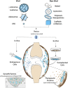
FIGURE 1
FIGURE 1
Intra-articular gene transfer- an archetypical model for orthopaedic gene therapy. (Top) Experimental strategies for delivery of therapeutic genes and nucleic acids to diseased or damaged tissues involve a variety of Viral and Non-Viral vector systems. The graphic on the left illustrates the basic structural and physical properties of the most widely used viral vector systems. The Retroviruses (γ-retrovirus and lentivirus) are relatively large (∼100 nm dia) enveloped viruses that “bud” from the surface of the infected cell. The outer envelope is composed of lipid bilayer acquired from the plasma membrane of the host cell during viral escape. Retroviral env (or VSV-G for pseudotyped virus) glycoprotein molecules transverse the outer envelope and are used for viral attachment to target cells. Two + strand copies of the RNA genome are encased in the protective nucleocapsid along with reverse transcriptase and integrase proteins that convert the RNA genome to DNA and integrate the provirus into the host genome. Adenovirus is a non-enveloped virus that replicates by lytic infection. The viral capsid is an icosahedron ∼90–100 nm in diameter that encases a linear dsDNA genome. Adenoviral fiber/knob complexes protrude from each of the 12 vertices of the icosahedron and are used for attachment to target cells. Following entry into the nucleus, the viral DNA remains episomal. Adeno-associated virus (AAV) is a comparatively simple, small non-enveloped, iscosahedral virus, ∼20 nm in diameter. The viral capsid houses a short (∼4.7 bp) ssDNA genome. Following infection, the genome is maintained episomally in concatemers formed by intermolecular recombination (Yang et al., 1999). Due to their inherent differences in biology and physical properties each viral vector is best suited to different types of applications and delivery strategies. To bypass the need for viruses for gene delivery, a wide range of non-viral systems (shown in the graphic on the right) are under investigation for their utility in orthopaedic applications. These non-viral systems utilize to varying extents, chemical modification of plasmid DNAs, soluble mRNAs, and RNAi molecules, which can be delivered “naked” in soluble form, or complexed with cationic lipids as liposomes or various polymers into nanoparticles to condense and protect the nucleic acids from degradation, prevent electrostatic repulsion and facilitate cellular uptake. Various carrier scaffolds and matrices are often employed to aid and prolong delivery to target cells. (Bottom) Once incorporated in an appropriate vector, the therapeutic gene or nucleic acid can be delivered to diseased or damaged tissues by either in vivo or ex vivo methods. For in vivo delivery, the vector is administered directly to tissues at the relevant site to modify the resident cell populations in situ. In the present example, the vector is injected intra-articularly into the synovial fluid of an arthritic joint to diffuse throughout the joint cavity and modify endogenous cells in the synovial lining (shown in blue) and/or articular cartilage. For ex vivo delivery, the vector is used to modify cells growing in culture, which can be administered locally to the site of disease or injury by different routes depending on the application. As indicated by the black arrow, the modified cells can be injected into the joint (or other relevant site) as a cellular suspension, to disperse and engraft in the local tissues to continuously express and secrete a therapeutic gene product (e.g., IL-1Ra, IL-10 etc.) into the local fluids and tissues to inhibit inflammatory signaling for an extended duration (right-hand inset). Alternatively, the modified cells can be incorporated into a support matrix and surgically implanted into a focal cartilage lesion (or other damaged tissue) (dashed gray arrow). Following delivery, the modified cells continually release specific growth factors to stimulate chondrogenic differentiation and cartilage matrix synthesis to facilitate repair by both the local and implanted cell populations (left-hand inset).
FIGURE 2
FIGURE 2
Organization of the cis-acting sequence elements in a typical expression cassette for therapeutic gene transfer. For most vector systems, both viral and non-viral, the expression cassette is designed to provide high-level synthesis of the transgene product independent of the regulatory constraints of the endogenous gene. The specific sequences of the elements in a particular cassette are often assembled from a variety of sources, both eukaryotic and viral (Keravala and Gasmi, 2021). The Promoter located at the 5′ end of the cassette, drives transcription of the therapeutic transgene (often the cDNA of a secreted protein). The transcription start site and direction of RNA synthesis are indicated by the arrow. The DNA sequences downstream from the promoter serve as the template for RNA synthesis, and the regions indicated represent the cis- acting RNA sequences in the 5′ and 3′ untranslated regions (UTRs) to enhance or regulate translation of the RNA transcript. The Intron: as a cDNA represents the protein coding sequence of a mature mRNA, an intronic sequence with flanking splice donor (SD) and acceptor (SA) sites is used to direct splicing of the primary transcript to enhance nuclear export and translation. The cDNA: the locations and template sequences of the translation start and stop codons are shown in bold. A consensus Kozac sequence flanks the ATG start codon, and during codon optimization is engineered into the sequence of the cDNA to enhance translation initiation and prevent cryptic starts at internal ATG (AUG) codons. miRNA Binding Sites: in the 3′ UTR, recognition sites for the binding regions of select miRNAs can be inserted to fine-tune or conditionally modulate mRNA translation in specific applications. WPRE: a woodchuck hepatitis virus post-transcriptional regulatory element or similar PRE can be inserted to enhance nuclear export and translation. Poly A: the polyadenylation signal at the 3′ end of the transcript serves as a cleavage site for the addition of the polyadenosine tract, which promotes nuclear export, mRNA stability and translation. Scissors: designate cloning sites for removal or insertion of cDNA(s) of interest. Regions internal to the cloning sites represent sequence elements specific to individual applications, while those outside are more generic and stably reside in the expression cassette of the vector.
FIGURE 3
FIGURE 3
Promoter elements commonly used to drive therapeutic gene expression. Strong Constitutive Promoters: to compensate for limitations with gene delivery, most vector systems employ promoters with high basal activation for continuous high-level transgene expression. The diagram illustrates the differences in size and sequence components among some of the more widely used constitutive promoters. (The dark arrows indicate the transcription start site (TSS) and direction of RNA synthesis.) 1) the eukaryotic translation elongation factor 1α (EF1α) promoter: a short core promoter (CP) sequence lies upstream of the TSS with additional activation sequences located downstream in intron 1 of the EF1a gene (Wakabayashi-Ito and Nagata, 1994; Gopalkrishnan et al., 1999); 2) the cytomegalovirus (CMV) immediate early promoter/enhancer: composed of a minimal promoter (MP) that signals the TSS with short proximal and distal enhancer elements (PE and DE) immediately upstream (Prösch et al., 1996; Isomura et al., 2004) and 3) the early promoter from Simian Virus 40 (SV40): composed of a small minimal promoter with activation signals located in tandem 21 bp and 72 bp repeat sequences immediately upstream (Gendra et al., 2007). Other strong promoters common in the literature are Hybrids assembled from sequence elements of both eukaryotic and viral origin. Among these are 1) the chicken β-actin promoter (CBA/CAG) comprised of the CMV distal enhancer (DE) positioned upstream of the CBA core promoter, followed by the splice donor and enhancer elements from CBA intron 1 fused to the splice acceptor site of exon 3 of the rabbit b-globin gene (rβg) (Xu et al., 2001; You et al., 2010), and 2) the derivative CBh promoter composed of the CMV DE and CBA core promoter, with a hybrid intron immediately downstream composed of the splice donor site from CBA intron 1 fused to the splice acceptor site from minute virus of mice (MVM) VP intron (Gray et al., 2011). As each can provide high-level expression in the proper contexts, promoter selection is heavily influenced by size- of both the promoter and transgene and available space within vector. Inducible Promoters: for certain applications where conditional expression is desired, a wide range of synthetic inducible promoter systems are commercially available, or can be readily constructed/synthesized using the minimal CMV promoter as the transcription start site linked to an upstream array of up to 30 response/recognition elements (REs) (Ede et al., 2016) for potent transcription factor(s) induced by a particular change(s) in growth conditions, such as NF-kB (inflammation) or HIF1 (hypoxia) (Shibata et al., 2000). Alternatively transgene expression can be induced externally by the presence of a chemical agent, such as tetracycline which enables a Tet-binding transactivator (Tet/ta) to bind and interact with the TetO cis element (Loew et al., 2010). Tissue Specific Promoters: to prevent toxicity from transgene expression in off-target tissues, a number of tissue-specific promoters have been developed using the endogenous regulatory sequences. These elements are typically large, with weak transgene expression relative to ubiquitously active promoters, e.g., the muscle-specific desmin promoter/enhancer (DES) (Talbot et al., 2010). The muscle creatine kinase (CMK), Muscle Hybrid (MH) promoter designed in silico from muscle-specific transcription factor binding clusters is comprised of the desmin and CMK enhancer regions upstream of the CMK core promoter and followed downstream by a small intronic enhancer element (SIE) (Piekarowicz et al., 2019). In vitro and in vivo gene expression from the MH promoter was 2–4x that of CMV and >100x greater than the desmin promoter. A small liver-specific hybrid promoter comprised of core promoter for human α1 antitrypsin (hAAT) linked to upstream apolipoprotein E enhancer elements provides potent transgene expression in hepatocyte cultures and in liver, equivalent to CMV (Gehrke et al., 2003).
FIGURE 4
FIGURE 4
Comparison of the internal anatomic structures of the stifle joints (hind knees) of the adult rat (inset) and the horse, which is comparable in size to the human knee. The striking differences in magnitude, volume and thickness of the tissues underscore the practical challenges associated with the scale-up of a gene-based therapy to a large mammalian system.
FIGURE 5
FIGURE 5
GFP expression in healthy and OA joints following intra-articular gene delivery with scAAV. The middle carpal joints of 3 healthy horses and 3 with late stage naturally-occurring OA were injected with 5 × 10¹² vg of scAAV.GFP. Two weeks later the joint tissues were collected and analyzed for fluorescence. (A) (Top row) Fluorescence activity in freshly harvested synovial tissues viewed with inverted fluorescence microscopy at ×10 magnification. (bottom row) Paraffin sections of synovium immunohistochemically stained for GFP at ×20 magnification. In normal joints, the synovium was the predominant site of transgene expression, with abundant fluorescent cells scattered throughout the capsular lining, often concentrated in thicker villous regions. In striking contrast, the number and density of the fluorescent cells in OA joints were visibly greater across the entire expanse of the synovial lining, but particularly so in regions with marked hyperplasia and leukocytic infiltration. In both normal and OA joints the transduced cells were almost exclusively delimited to the synovium and subsynovium, and only rarely seen in the supporting fibrous tissues. (B) GFP expression in fresh cartilage shavings viewed with inverted fluorescence microscopy. Images in the top and bottom rows are at ×10 and ×20 magnification, respectively. In articular cartilage from normal joints, GFP fluorescence was visible but generally faint and limited to scattered isolated cells. In OA cartilage, GFP activity was dramatically enhanced, as populations of brightly fluorescent cells were readily apparent in all shavings recovered. The labeled chondrocytes included both elongated cells, consistent with superficial layer chondrocytes, and cells with more spherical morphology characteristic of chondrocytes in deeper layers. Shavings harvested near full thickness erosions often contained focal regions with intense fluorescence readily visible at low magnification.
FIGURE 6
FIGURE 6
Severe synovial fibrosis and chondrometaplasia induced by intra-articular gene delivery of TGF-β1. The stifle joints (hind knees) of nude rats were injected bilaterally with 2 × 10⁹ viral particles of Ad.TGF-β1. Groups of animals were killed at days 0, 5, 10, and 30, and the joints were harvested and processed for histology. Adjacent sections were stained with H&E or toluidine blue as indicated. The images in left two columns are at ×2.5 magnification, while those in the right two columns are at ×20. At Day 5, expansion of spindled fibroblasts from the synovial lining and joint capsule produced a dense fibrotic mass that fully occluded the underlying adipose layer. By Day 10, the fibrotic tissue had expanded to displace all the soft tissue structures and began to fuse with articular cartilage, in which the development of rounded chondrocytic cells can be seen. By Day 30, the bulk of the fibrotic expanse had differentiated into a cartilaginous phenotype as indicated by the cellular morphology and metachromatic toluidine blue staining. In some areas the normal articular cartilage was replaced by metaplastic fibrocartilaginous tissue that also permeated the subchondral and periarticular bone. Figure copied with permission from (Watson et al., 2010).

References

    1. Aghaloo T., Jiang X., Soo C., Zhang Z., Zhang X., Hu J., et al. (2007). A Study of the Role of Nell-1 Gene Modified Goat Bone Marrow Stromal Cells in Promoting New Bone Formation. Mol. Ther. 15, 1872–1880. 10.1038/sj.mt.6300270 PubMed Abstract | 10.1038/sj.mt.6300270 | Google Scholar - DOI - DOI - PMC - PubMed
    1. Alaee F., Bartholomae C., Sugiyama O., Virk M., Drissi H., Wu Q., et al. (2015). Biodistribution of LV-TSTA Transduced Rat Bone Marrow Cells Used for "Ex-Vivo" Regional Gene Therapy for Bone Repair. Cgt 15, 481–491. 10.2174/1566523215666150812120229 10.2174/1566523215666150812120229 | Google Scholar - DOI - DOI - PubMed
    1. Alba R., Bosch A., Chillon M. (2005). Gutless Adenovirus: Last-Generation Adenovirus for Gene Therapy. Gene Ther. 12 (Suppl. 1), S18–S27. 10.1038/sj.gt.3302612 PubMed Abstract | 10.1038/sj.gt.3302612 | Google Scholar - DOI - DOI - PubMed
    1. Alluri R., Song X., Bougioukli S., Pannell W., Vakhshori V., Sugiyama O., et al. (2019). Regional Gene Therapy with 3D Printed Scaffolds to Heal Critical Sized Bone Defects in a Rat Model. J. Biomed. Mater Res. 107, 2174–2182. 10.1002/jbm.a.36727 10.1002/jbm.a.36727 | Google Scholar - DOI - DOI - PMC - PubMed
    1. Altawalah H. (2021). Antibody Responses to Natural SARS-CoV-2 Infection or after COVID-19 Vaccination. Vaccines (Basel) 9, 910. 10.3390/vaccines9080910 PubMed Abstract | 10.3390/vaccines9080910 | Google Scholar - DOI - DOI - PMC - PubMed