. 2021 Dec 13;11(3):219-223.
doi: 10.1055/s-0041-1735885. eCollection 2022 Jun.

Classification and Management of Failed Fixation of the Volar Marginal Fragment in Distal Radius Fractures

Affiliations

Classification and Management of Failed Fixation of the Volar Marginal Fragment in Distal Radius Fractures

M Carolina Orbay et al. J Wrist Surg. .

Abstract

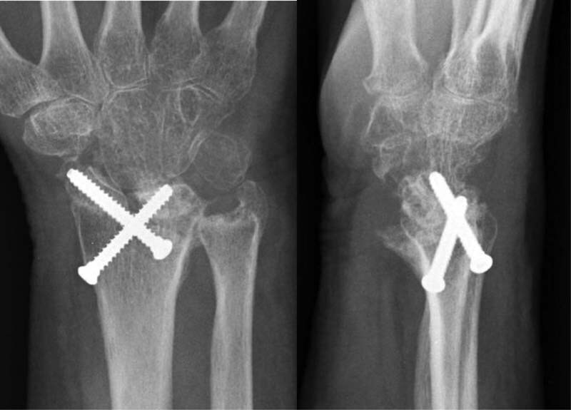
Greater understanding of specific fracture patterns following distal radius fractures has arisen with the advent of volar plating. The volar marginal fragment (VMF) is a small peripheral piece of bone which is critical to carpal stability. Failure to achieve good fixation of the VMF can result in volar subluxation of the carpus and distal radioulnar joint instability. Due to its small, distal nature, this fragment can be easily missed and difficult to fix. Loss of reduction of the VMF following operative fixation presents specific challenges and surgical considerations dictated by patient characteristics and timing. Our goal of this review is to present a classification system for these failed VMFs which can help guide surgical treatment as well as expected outcomes.

Keywords: distal radius fractures; hook plate; revision distal radius; volar lunate facet; volar marginal fragment.


Conflict of interest statement

Conflict of Interest J.L.O. is a consultant for Skeletal Dynamics. J.L.O. is the first inventor on the Geminus Volar Plating System patent. J.L.O. certifies that he may receive payments or benefits from Skeletal Dynamics.

Figures

Fig. 1
Fig. 1
Anteroposterior and lateral radiographs of a patient who presented 2 weeks following attempted fixation of a complex fracture with a standard volar locked plate and Kirschner wires. Failure of fixation is evident on the lateral radiograph (arrow) with a “tear drop angle” greater than 70 degrees and the carpus subluxated. Subluxation is confirmed, as the center of the lunate is palmar to the volar radial cortex (longitudinal line).
Fig. 2
Fig. 2
Anteroposterior and lateral radiographs of revision fixation of the failed volar marginal fragment (VMF). The VMF was captured volarly using an extension hook plate. Dorsal bridge plate fixation neutralized the joint forces and maintained reduction of the carpus.
Fig. 3
Fig. 3
Lateral radiograph taken 3 months after fixation of distal radius fracture with volar marginal fragment. Resorption of the fracture fragment has resulted in subchondral collapse, volar subluxation of the carpus, and articulation of the lunate against the plate (circle).
Fig. 4
Fig. 4
Anteroposterior and lateral radiographs taken 5 months after volar opening wedge osteotomy. Note the volar plate was bent to deliver neutral tilt. Autologous corticocancellous bone graft was used to support the volar rim and fill the bony void below the lunate facet. Patient maintained functional range of motion and had significant improvement of pain following intervention. Consideration should be given to subsequent plate removal.
Fig. 5
Fig. 5
Multiple combined techniques: volar tilt corrected to neutral with a volar opening wedge osteotomy and supported with corticocancellous autograft. An extension hook plate used to capture the volar marginal fragment. A dorsal bridge plate used to unload the articular surface and aid with radiocarpal reduction. The lunate was temporarily pinned to the radius with Kirschner wires.
Fig. 6
Fig. 6
Anteroposterior and lateral radiographs taken 6 months following proximal row to radius fusion. The relationship between the scaphoid and the lunate was preserved by not interrupting the scapholunate ligament and initially preserving the interval between these two carpal bones. The radial and proximal chondral surfaces of the scaphoid and lunate are partially decorticated with a small burr following fixation and grafted with autologous graft obtained from the distal radius.

Similar articles

Cited by

References

    1. Majima M, Horii E, Matsuki H, Hirata H, Genda E. Load transmission through the wrist in the extended position. J Hand Surg Am. 2008;33(02):182–188. - PubMed
    1. Benis S, Vanhove W, Hollevoet N. Volar plate fixation in intra-articular distal radius fractures with a volar lunate facet fragment. Hand Surg Rehabil. 2020;39(04):270–274. - PubMed
    1. Naito K, Sugiyama Y, Kinoshita M. Functional outcomes in volar-displaced distal radius fractures patients with marginal rim fragment treated by volar distal locking plates. J Hand Microsurg. 2019;11(02):100–105. - PMC - PubMed
    1. O'Shaughnessy M A, Shin A Y, Kakar S. Volar marginal rim fracture fixation with volar fragment-specific hook plate fixation. J Hand Surg Am. 2015;40(08):1563–1570. - PubMed
    1. Orbay J L, Rubio F, Vernon L L. Prevent collapse and salvage failures of the volar rim of the distal radius. J Wrist Surg. 2016;5(01):17–21. - PMC - PubMed