. 2022 Jul 15;132(14):e157262.
doi: 10.1172/JCI157262.

CD11b suppresses TLR activation of nonclassical monocytes to reduce primary graft dysfunction after lung transplantation

Affiliations

CD11b suppresses TLR activation of nonclassical monocytes to reduce primary graft dysfunction after lung transplantation

Melissa Querrey et al. J Clin Invest. .

Abstract

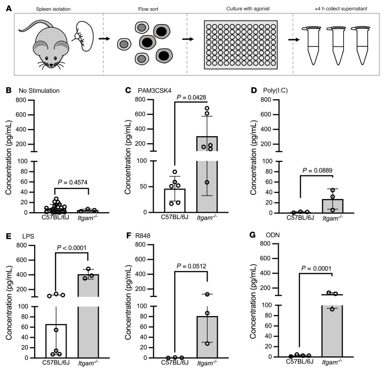
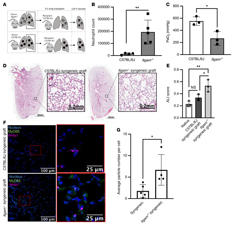
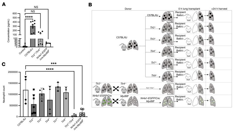
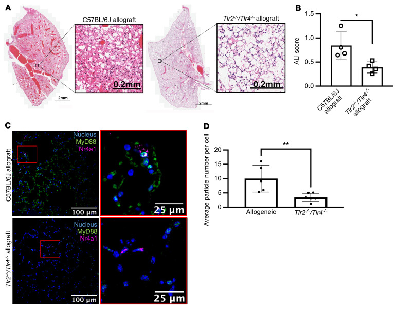
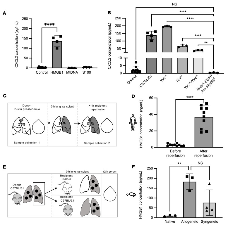
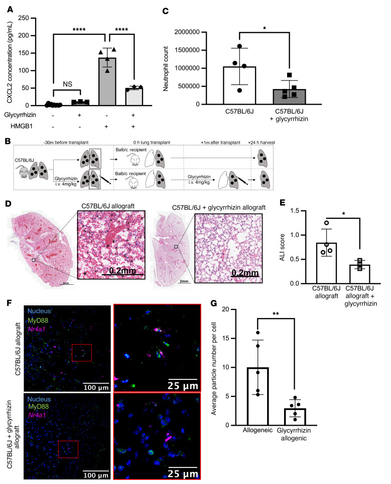
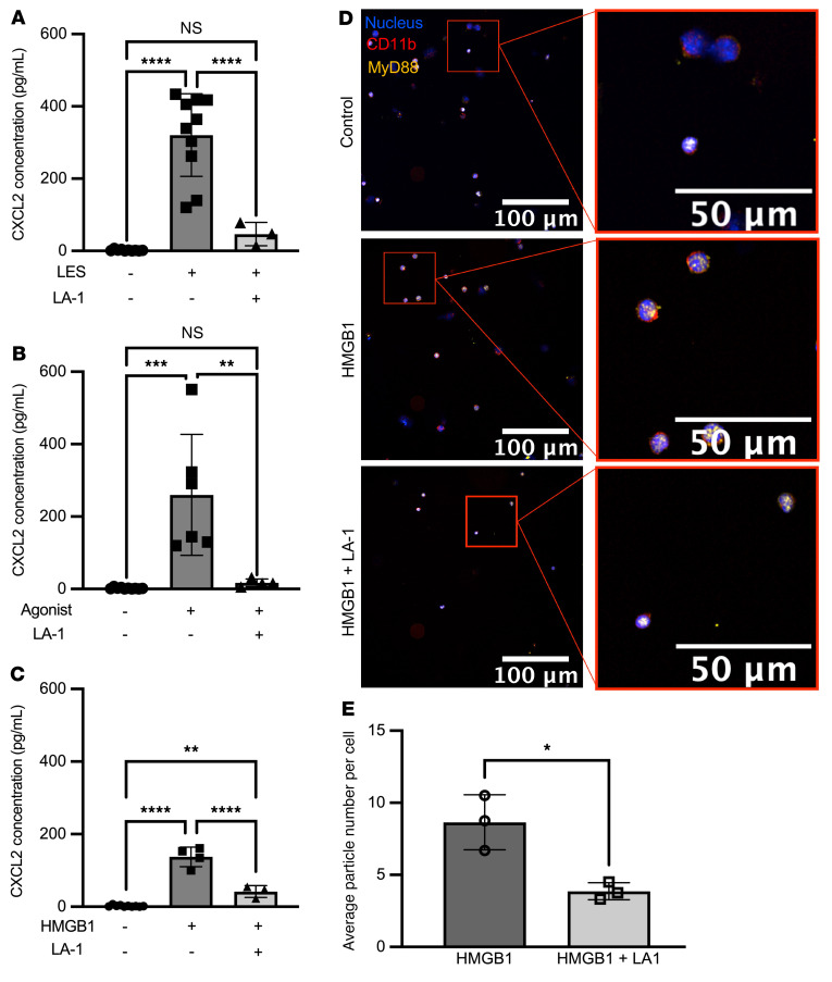
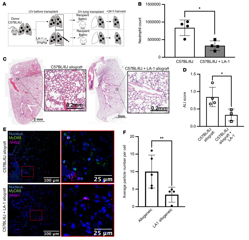
Primary graft dysfunction (PGD) is the leading cause of postoperative mortality in lung transplant recipients and the most important risk factor for development of chronic lung allograft dysfunction. The mechanistic basis for the variability in the incidence and severity of PGD between lung transplant recipients is not known. Using a murine orthotopic vascularized lung transplant model, we found that redundant activation of Toll-like receptors 2 and 4 (TLR2 and -4) on nonclassical monocytes activates MyD88, inducing the release of the neutrophil attractant chemokine CXCL2. Deletion of Itgam (encodes CD11b) in nonclassical monocytes enhanced their production of CXCL2 and worsened PGD, while a CD11b agonist, leukadherin-1, administered only to the donor lung prior to lung transplantation, abrogated CXCL2 production and PGD. The damage-associated molecular pattern molecule HMGB1 was increased in peripheral blood samples from patients undergoing lung transplantation after reperfusion and induced CXCL2 production in nonclassical monocytes via TLR4/MyD88. An inhibitor of HMGB1 administered to the donor and recipient prior to lung transplantation attenuated PGD. Our findings suggest that CD11b acts as a molecular brake to prevent neutrophil recruitment by nonclassical monocytes following lung transplantation, revealing an attractive therapeutic target in the donor lung to prevent PGD in lung transplant recipients.

Keywords: Immunology; Integrins; Monocytes; Transplantation.


Conflict of interest statement

Conflict of interest: The authors have declared that no conflict of interest exists.

Figures

Figure 1
Figure 1. Itgam–/– increases CXCL2 production in response to TLR2 and TLR4 agonists.
(A) Splenic NCMs were flow cytometry sorted from wild-type (C57BL/6J) and Itgam–/– mice and stimulated with TLR agonists as indicated for 4 hours, after which the supernatants were collected for ELISA analysis. (BG) PAM3CSK4, a TLR2 agonist; poly(I:C), a TLR3 agonist; lipopolysaccharide (LPS), a TLR4 agonist; resiquimod (R848), a TLR 7/8 agonist; and oligodeoxynucleotide (ODN), a TLR9 agonist were administered at a dose of 1 μg. Each symbol represents 50,000 NCMs, and approximately 100,000–200,000 NCMs were isolated from an individual mouse. P values above the horizontal lines were calculated using a 2-tailed, unpaired t test.
Figure 2
Figure 2. Itgam–/– donor lungs exacerbate development of PGD.
(A) Schematic for murine syngeneic lung transplants. (B) Lung allografts were harvested 24 hours after lung transplantation and neutrophil numbers were measured using flow cytometry. **P = 0.0019 by 2-tailed, unpaired t test. (C) Arterial blood was obtained for blood gas analysis while the mouse was receiving 100% oxygen via mechanical ventilation immediately prior to harvest. *P = 0.0206 by 2-tailed, unpaired t test. (D) Representative H&E-stained allografts from wild-type and Itgam–/– mice. (E) Acute lung injury (ALI) scores based on histologic evaluation. *P = 0.0329, **P = 0.0046 by 1-way ANOVA with Dunnett’s test to correct for multiple comparisons. NS, not significant. (F) Lung sections from allografts of wild-type and Itgam–/– mice were stained using a combination of single-molecule fluorescence in situ hybridization (RNAScope) and immunohistochemistry (blue: nuclear stain, green: MyD88, magenta: Nr4a1). Original magnification, ×400. (G) MyD88-containing aggregates per Nr4a1-positive NCM in C57BL/6J and Itgam–/– syngeneic grafts. *P = 0.0261 by 2-tailed, unpaired t test. Each symbol represents an individual mouse.
Figure 3
Figure 3. Activation of TLR2 or TLR4 is necessary for CXCL2 release from NCMs and the development of PGD after murine lung transplantation.
(A) CXCL2 in supernatants from flow cytometry–sorted splenic NCMs from C57BL/6J, Tlr2–/– Tlr4–/–, and Nr4a1-EGFP/cre Myd88fl/fl mice after stimulation with lysed endothelial supernatant for 4 hours. ****P < 0.0001 by 1-way ANOVA with Dunnett’s correction for multiple comparisons. (B) Schematic for the allogeneic transplantations in C. Donor mice were C57BL/6J (wild type), Tlr2–/–, Tlr3–/–, Tlr4–/–, Tlr7–/–, Tlr9–/–, Tlr2–/– Tlr4–/–, and Nr4a1-EGFP/cre Myd88fl/fl. All recipients were BALB/c mice. Lungs were harvested 24 hours after transplantation. (C) Neutrophils were quantified by flow cytometry analysis of lung homogenates. ***P = 0.0006, ****P < 0.0001 by 1-way ANOVA with Dunnett’s multiple comparison test, as detailed in Supplemental Figure 5. NS, not significant. Each symbol represents an individual mouse.
Figure 4
Figure 4. Activation of TLR2 or TLR4 is necessary for the development of PGD after murine lung transplantation.
(A) Representative H&E staining of wild-type and Tlr2–/– Tlr4–/– allografts show less necrosis and inflammatory infiltrate with preservation of lung structure. (B) Acute lung injury (ALI) scores based on histologic evaluation. *P = 0.0162 by 1-way ANOVA with Dunnett’s multiple comparisons test, as detailed in Supplemental Figure 5. (C) Lung sections from wild-type and Tlr2–/– Tlr4–/– mice were analyzed using a combination of RNAScope and immunohistochemical staining of allografts (blue: nuclear stain, green: MyD88, magenta: Nr4a1). Original magnification, ×400. (D) MyD88-containing aggregates per Nr4a1-positive NCM in C57BL/6J and Tlr2–/– Tlr4–/– allografts. **P = 0.0032 by 1-way ANOVA with Dunnett’s multiple comparisons test, as detailed in Supplemental Figure 5. Each symbol represents an individual mouse. The controls for ALI score and average particle number per cell are identical to those in Figures 6 and 8.
Figure 5
Figure 5. HMGB1 activates TLR2 and TLR4 to induce the expression of CXCL2 in NCMs.
(A) Splenic NCMs were flow cytometry sorted from wild-type (C57BL/6J) mice and stimulated with putative DAMPs as indicated for 4 hours, after which the supernatants were collected for ELISA analysis. ****P < 0.0001 by 1-way ANOVA with Tukey’s multiple comparisons test. Mitochondrial DNA (mtDNA), S100 proteins (S100), and HMGB1 were administered at a dose of 1 μg. (B) Splenic NCMs were flow cytometry sorted from wild-type (C57BL/6J), Tlr2–/–, Tlr4–/–, and Tlr2–/– Tlr4–/– mice and treated with HMGB1 (1 μg), and CXCL2 was measured in the supernatant 4 hours later. **P = 0.0022, ****P < 0.0001 by 1-way ANOVA with Tukey’s multiple comparisons test. NS, not significant. (C) Schematic for human sera collection in D. (D) Blood samples from patients undergoing lung transplantation were obtained immediately prior to and 24 hours after reperfusion and analyzed for HMGB1. ****P < 0.0001 by 2-tailed, paired t test. (E) Schematic for murine sera collection. (F) HMGB1 serum concentrations in native, allogeneic, and syngeneic lung transplants. **P = 0.0073 by 1-way ANOVA with Dunnett’s correction for multiple comparisons. In A and B, each symbol represents 50,000 NCMs, and approximately 100,000 to 200,000 NCMs were isolated from an individual mouse; each symbol in D and F represents an individual mouse or human.
Figure 6
Figure 6. Glycyrrhizin inhibits HMGB1 activation of NCMs and prevents PGD in mice.
(A) Splenic NCMs were flow cytometry sorted from wild-type (C57BL/6J) mice and treated with glycyrrhizin (20 μM) followed 20 minutes later by HMGB1 (10 μg/mL), and CXCL2 concentrations in the supernatants were measured 4 hours later. ****P < 0.0001 by 1-way ANOVA with Tukey’s multiple comparisons test. (B) Schematic for the allogeneic transplantations in CE. Donor mice were C57BL/6J and recipients were BALB/c. Glycyrrhizin was administered at 4 mg/kg intravenously to the donor 30 minutes before harvest and to the recipient immediately after reperfusion. (C) Lung allografts were harvested 24 hours after lung transplantation and neutrophil numbers were quantified by flow cytometry analysis of lung homogenates. *P = 0.0235 by 1-way ANOVA with Dunnett’s correction for multiple comparisons, as detailed in Supplemental Figure 5. Each symbol represents an individual mouse. (D) Representative H&E staining of allografts with and without glycyrrhizin treatment. (E) Acute lung injury (ALI) scores for the images in D. *P = 0.0259 by 1-way ANOVA with Dunnett’s correction for multiple comparisons (see Supplemental Figure 5). (F) Lung sections from allografts after syngeneic lung transplantation with or without treatment with glycyrrhizin using RNAScope and immunohistochemical staining (blue: nuclear stain, green: MyD88, magenta: Nr4a1). Original magnification, ×400. (G) MyD88 particle counts per Nr4a1-positive NCM in C57BL/6J allografts with and without glycyrrhizin treatment. **P = 0.0025 by 1-way ANOVA with Dunnett’s multiple comparisons test, as detailed in Supplemental Figure 5. Each symbol represents 50,000 NCMs. The controls for ALI score and average particle number per cell are identical to those in Figures 4 and 8.
Figure 7
Figure 7. LA-1 decreases CXCL2 production in NCMs.
(AC) Splenic NCMs were flow cytometry sorted from wild-type (C57BL/6J) mice and then treated with leukadherin-1 (LA-1) (20 μM) 15 minutes prior to the indicated TLR agonists, and CXCL2 was measured in the supernatant 4 hours later. PAM3CSK4, LPS, lysed endothelial supernatant (LES), and HMGB1 were administered at a dose of 10 μg/mL. **P = 0.0019 (B); P = 0.0020 (C); ***P = 0.0001; ****P < 0.0001 by 1-way ANOVA with Tukey’s multiple comparisons test. NS, not significant. (D) Splenic NCMs from Nr4a1-EGFP mice were flow cytometry sorted and treated with LA-1 (20 μM) followed 15 minutes later by HMGB1 (10 μg/mL), and 4 hours later cytospins of these cells were stained for immunofluorescence analysis (blue: nuclear, green: CD11b, magenta: MyD88). Original magnification, ×400. (E) MyD88 particle count in isolated NCMs stimulated with HMGB1 with and without LA-1 treatment. *P = 0.0143 by 2-tailed, unpaired t test. Each symbol represents 50,000 NCMs, and approximately 100,000–200,000 NCMs were isolated from an individual mouse.
Figure 8
Figure 8. LA-1 prevents PGD in mice.
(A) Schematic for the allogeneic transplantations. Donor mice were C57BL/6J and recipients were BALB/c. LA-1 was administered (2 mg/kg intravenously) only to the donor. (B) Lung allografts were harvested 24 hours after lung transplantation and neutrophil numbers were quantified from lung homogenates using flow cytometry. *P = 0.0128 by 1-way ANOVA with Dunnett’s correction for multiple comparisons, as detailed in Supplemental Figure 5. (C) Representative H&E staining of allografts with and without glycyrrhizin treatment. (D) Acute lung injury (ALI) scores for the images in C. *P = 0.0122 by 1-way ANOVA with Dunnett’s correction for multiple comparisons, as detailed in Supplemental Figure 5. (E) Lung sections from allografts of mice with and without glycyrrhizin treatment using RNAscope and immunohistochemical staining (blue: nuclear stain, green: MyD88, magenta: Nr4a1). Original magnification, ×400. (F) MyD88 particle counts per Nr4a1-positive NCM in allografts with and without LA-1 treatment. **P = 0.0070 by 1-way ANOVA with Dunnett’s multiple comparisons test, as detailed in Supplemental Figure 5. Each symbol represents an individual mouse. The controls for ALI score and average particle number per cell are identical to those in Figures 4 and 6.

Similar articles

Cited by

References

    1. Suzuki Y, et al. Primary graft dysfunction. Sem Respir Crit Care Med. 2013;34(3):305–319. doi: 10.1055/s-0033-1348474. - DOI - PMC - PubMed
    1. Morrison MI, et al. Pathophysiology and classification of primary graft dysfunction after lung transplantation. J Thorac Dis. 2017;9(10):4084–4097. doi: 10.21037/jtd.2017.09.09. - DOI - PMC - PubMed
    1. Christie JD, et al. Primary graft failure following lung transplantation. Chest. 1998;114(1):51–60. doi: 10.1378/chest.114.1.51. - DOI - PubMed
    1. Lee JC, et al. Primary graft dysfunction: definition, risk factors, short- and long-term outcomes. Sem Respir Crit Care Med. 2010;31(2):161–171. doi: 10.1055/s-0030-1249111. - DOI - PubMed
    1. Shah RJ, Diamond JM. Primary graft dysfunction (PGD) following lung transplantation. Semin Rspir Crit Care Med. 2018;39(2):148–154. doi: 10.1055/s-0037-1615797. - DOI - PubMed

Publication types

Substances