. 2023;26(6):1204-1213.
doi: 10.2174/1386207325666220715090505.

Penehyclidine Hydrochloride Protects Rat Cardiomyocytes from Ischemia- Reperfusion Injury by Platelet-derived Growth Factor-B

Affiliations

Penehyclidine Hydrochloride Protects Rat Cardiomyocytes from Ischemia- Reperfusion Injury by Platelet-derived Growth Factor-B

Yan Lu et al. Comb Chem High Throughput Screen. 2023.

Abstract

Aims and objective: The lack of effective treatments for myocardial ischemiareperfusion (MI-R) injury severely restricts the effectiveness of the treatment of ischemic heart disease. In the present research, we aimed to investigate the protective effect and molecular mechanism of penehyclidine hydrochloride (PHC) on MI-R cells.

Methods: Cell viability was quantified using CCK8. Cell apoptosis was analyzed using flow cytometry. Western blot and Elisa assays were used for the detection of target proteins.

Results: PHC pretreatment attenuated the inhibition of cell viability and decreased the percentage of apoptosis induced by simulated ischemia reperfusion (SIR). Platelet-derived growth factor B (PDGF-B) and its downstream AKT pathway were activated in PHC pretreated cells. After siRNAPDGF- B transfection, cell viability was inhibited and apoptosis was activated in PHC pretreated SIR cells, suggesting that PHC protected cells from SIR. PDGF-B knockdown also increased the levels of CK, LDH, IL-6 and TNF-α in PHC pretreated SIR cells. The effect of AKT inhibitor on H9C2 cells was consistent with that of PDGF-B knockdown.

Conclusion: PHC pretreatment can protect cardiomyocytes from the decrease of cell activity and the increase of apoptosis caused by reperfusion through up-regulating PDGF-B to activate PI3K pathway. Our study indicates that PHC is a potential drug to protect cells from reperfusion injury and PDGF-B is a potential target for preventing MI-R injury.

Keywords: Apoptosis; H9C2; cell viability; inflammatory factors; myocardial ischemia-reperfusion injury; penehyclidine hydrochloride.


Conflict of interest statement

The authors declare no conflict of interest financial or otherwise.

Figures

Fig. (1)
Fig. (1)
PHC protects H9C2 cells from reperfusion injury. (A), H9C2 cells of simulated ischemia reperfusion (SIR) group were incubated in DMEM with 95% N2/5% CO2 at 37°C for 6 h. Cells were pretreated with PHC (0, 0.025, 0.05, 0.1, 0.2, 0.5, 1, 5, 10 μM) for 2 h before SIR. Cell viability was detected using CCK8 assay. (B), cell micrographs of each group were taken after the incubation in DMEM with 95% N2/5% CO2 at 37°C for 6 h. (C) and (D), the levels of CK and LDH were detected using Elisa assay. (E), Flow cytometry was performed to quantify cell apoptosis. (F), the percentage of apoptotic cells was analyzed using FlowJo software. *P<0.05 vs. NC; #P<0.05 vs. SIR.
Fig. (2)
Fig. (2)
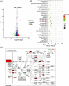
The potential targets and pathways by which PHC protects cells from reperfusion injury were analyzed through sequencing project. (A), Cells pretreated with 0.1 μM PHC for 2 h before SIR (SIR&PHC group) were sequencing with the SIR cells as the control. Differentially expressed genes between these two group cells were screened and a volcano plot was generated. (B), KEGG Pathway Enrichment of differentially expressed genes was performed using KOBAS (http://kobas.cbi.pku.edu.cn/). (C), Differentially expressed genes in PI3K-AKT signaling pathway. The original image KEGG graph was generated from KEGG (www.kegg.jp).
Fig. (3)
Fig. (3)
PHC promotes cell viability through PDGF-B and AKT pathway. (A), SIR&PHC cells were transfected with siRNA-PDGF-B to generate PHC&PDGF-B KD group, and were treated with 200 nmol/L BEZ235 to generate PHC&BEZ235 group. Western blot was performed to detect the phosphorylation levels of AKT and mTOR. (B) and (C), relative levels of p-AKT/AKT and p-mTOR/mTOR were measured using ImageJ software. *P<0.05 vs. SIR; #P<0.05 vs. SIR&PHC. (D), CCK8 was used to detect cell viability. *P<0.05; ***P<0.001.
Fig. (4)
Fig. (4)
PHC inhibits cell apoptosis through PDGF-B and AKT pathway. (A), Flow cytometry was performed to quantify cell apoptosis. (B), the percentage of apoptosis cells was analyzed using FlowJo software. (C), the expression levels of apoptosis-related proteins were detected using western blot. The relative levels of Bcl-2 (D), Bax (E), Casepase3-p17 (F) and Caspase3 (G) were quantified using ImageJ software. *P<0.05 vs. SIR; #P<0.05 vs. SIR&PHC.
Fig. (5)
Fig. (5)
PHC inhibits the expression levels of inflammatory factors through PDGF-B and AKT pathway. Elisa assay was performed to detect the levels of inflammatory factors. The relative levels of CK (A), LDH (B), IL-6 (C), IL-10 (D) and TNF-α (E) were analyzed. *P<0.05 vs. SIR; #P<0.05 vs. SIR&PHC.

Similar articles

Cited by

References

    1. Peng H., Abdel-Latif A. Cellular therapy for ischemic heart disease: An update. Adv. Exp. Med. Biol. 2019;1201:195–213. doi: 10.1007/978-3-030-31206-0_10. - DOI - PubMed
    1. Mokhtari-Zaer A., Marefati N., Atkin S.L., Butler A.E., Sahebkar A. The protective role of curcumin in myocardial ischemia-reperfusion injury. J. Cell. Physiol. 2018;234(1):214–222. doi: 10.1002/jcp.26848. - DOI - PubMed
    1. Kalogeris T., Baines C.P., Krenz M., Korthuis R.J. Ischemia/reperfusion. Compr. Physiol. 2016;7(1):113–170. doi: 10.1002/cphy.c160006. - DOI - PMC - PubMed
    1. Anzell A.R., Maizy R., Przyklenk K., Sanderson T.H. Mitochondrial quality control and disease: Insights into ischemia-reperfusion injury. Mol. Neurobiol. 2018;55(3):2547–2564. doi: 10.1007/s12035-017-0503-9. - DOI - PMC - PubMed
    1. Del Re D.P., Amgalan D., Linkermann A., Liu Q., Kitsis R.N. Fundamental mechanisms of regulated cell death and implications for heart disease. Physiol. Rev. 2019;99(4):1765–1817. doi: 10.1152/physrev.00022.2018. - DOI - PMC - PubMed

Publication types

MeSH terms

Substances