. 2022 Sep 16;23(18):e202200395.
doi: 10.1002/cbic.202200395. Epub 2022 Aug 16.

Cleavable Linker Incorporation into a Synthetic Dye-Nanobody-Fluorescent Protein Assembly: FRET, FLIM and STED Microscopy

Affiliations

Cleavable Linker Incorporation into a Synthetic Dye-Nanobody-Fluorescent Protein Assembly: FRET, FLIM and STED Microscopy

Ayse Aktalay et al. Chembiochem. .

Abstract

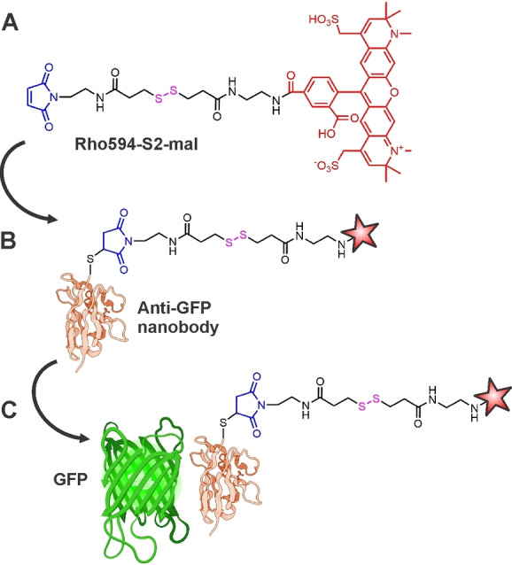
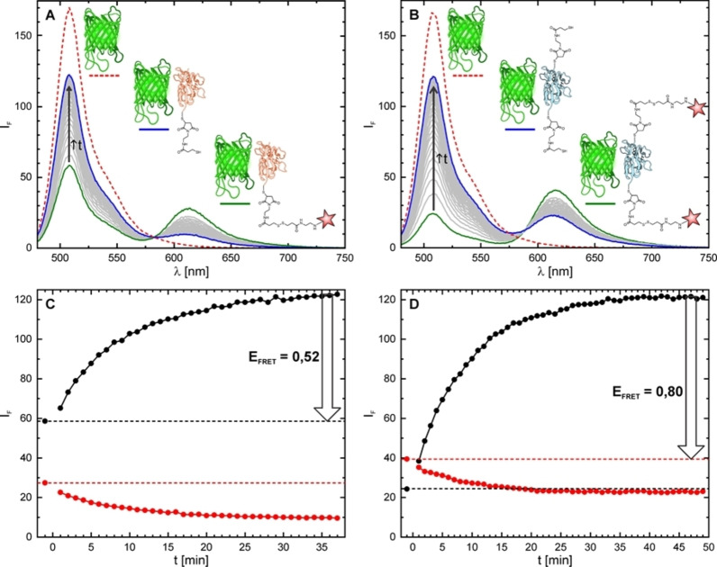
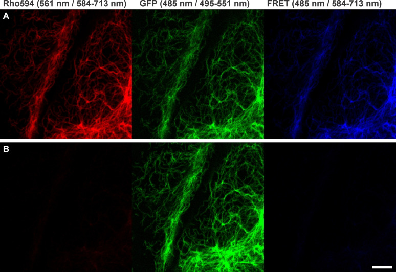
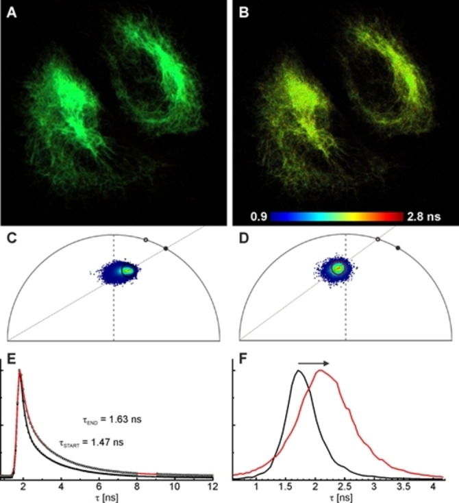
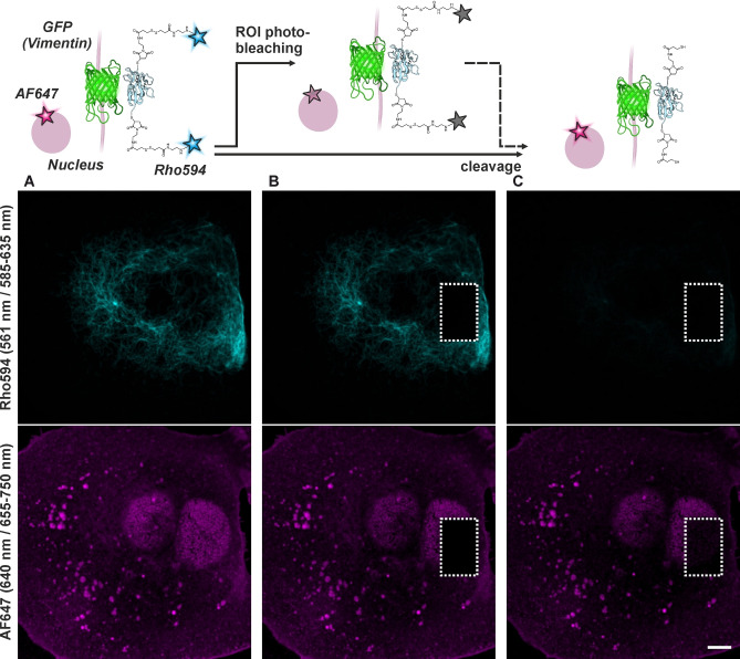
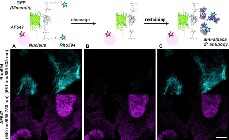
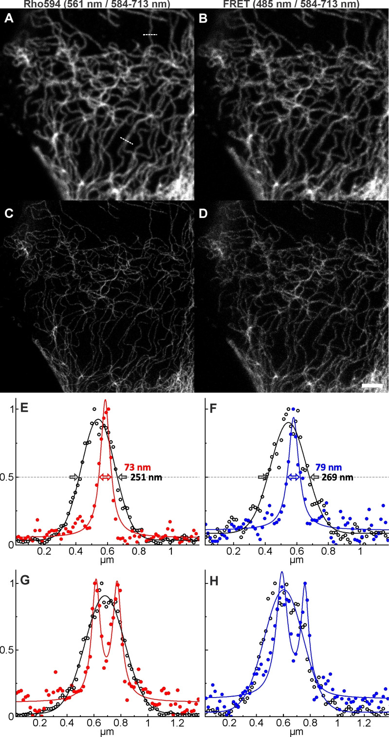
A bright and photostable fluorescent dye with a disulfide (S-S) linker and maleimide group (Rho594-S2-mal), as cleavable and reactive sites, was synthesized and conjugated with anti-GFP nanobodies (NB). The binding of EGFP (FRET donor) with anti-GFP NB labeled with one or two Rho594-S2-mal residues was studied in vitro and in cellulo. The linker was cleaved with dithiothreitol recovering the donor (FP) signal. The bioconjugates (FP-NB-dye) were applied in FRET-FLIM assays, confocal imaging, and superresolution STED microscopy.

Keywords: FRET; STED microscopy; dyes / pigments; fluorescence; nanobodies.


Conflict of interest statement

The authors declare no conflict of interest.

Figures

Figure 1
Figure 1
(A) Fluorescent probe Rho594‐S2‐mal incorporating the dye Alexa Fluor 594 (Rho594, shown in red), disulfide (S−S) linker (S2, black) and maleimide group (mal, blue) as cleavable and reactive sites, respectively. For synthesis, see Figures S1‐S2. (B) Anti‐GFP nanobody labelled with Rho594‐S2‐mal (another one with two cysteine residues was also labelled). (C) GFP (FRET donor) binds with the labelled anti‐GFP nanobody.
Figure 2
Figure 2
Emission spectra (top, ex. 470 nm, em. 480–750 nm), recorded in PBS (pH 7.4) at 25 °C, of EGFP alone (red) and EGFP in presence of Rho594‐S2‐mal labelled NBq (A) and NBx2 (C) before (green) and after (blue) cleavage of the dye by DTT. Concentration of EGFP and NB for all samples was 0.28 μM. Concentration of DTT was 5 mM. Evolution of fluorescence intensity over time (bottom) at 508 nm (black) and 612 nm (red) during cleavage from NBq (C) and NBx2 (D). DTT was added at t=0 min, and thus the first point corresponds to the protein bound with the nanobody (green line in A−B).
Figure 3
Figure 3
Confocal imaging with NBq‐Rho594‐S2‐mal on U2OS cells expressing rsEGFP2 on vimentin, before (A) and 1 h after (B) disulfide bond cleavage with DTT (5 mM). The emission signal was recorded in three different channels, for the acceptor, donor and FRET (donor excitation/acceptor emission) channel. The protein was activated with 405 nm irradiation on each pixel. Excitation wavelengths / detection windows are given on top. Scale bar: 5 μm.
Figure 4
Figure 4
Fluorescence lifetime imaging (FLIM) with NBx2‐(Rho594‐S2‐mal)2 on U2OS cells expressing rsEGFP2 on vimentin. Fast‐FLIM images before (A) and 1 h after (B) disulfide bond cleavage with DTT (1 mM), with the corresponding phasor plots (C−D). (E) Fluorescence decay of the FP emission before (filled symbols) and after bond cleavage (hollow symbols), along with biexponential fittings (black and red lines, respectively). (F) Fast‐FLIM histograms (mean arrival time) change during the reaction, as indicated by the arrow; the black and red curves correspond to the initial and final states in (A−E). Excitation: 488 nm, detection: 510–570 nm (donor channel). The protein was activated with 405 nm irradiation (with a CW laser) on each pixel.
Figure 5
Figure 5
Confocal imaging with NBx2‐(Rho594‐S2‐mal)2 on U2OS cells expressing rsEGFP2 on vimentin (cyan) and co‐stained with WGA/wheat germ agglutinine‐AF647 (magenta), before (A) and after acceptor depletion experiments by photobleaching only on the indicated ROI (B), and disulfide bond cleavage (C) with DTT (5 mM, rt, 1 h). Excitation wavelengths / detection windows are given on the left side (vertically). A scheme of consecutive steps is shown on top. Scale bar: 5 μm.
Figure 6
Figure 6
(A) Confocal imaging with NBx2‐(Rho594‐S2‐mal)2 on U2OS cells expressing rsEGFP2 on vimentin (cyan) and co‐stained with a primary antibody against NUP98 in combination with a secondary antibody labelled with AF647 (magenta). (B) Image acquired after disulfide bond cleavage with DTT (5 mM, rt, 1 h). (C) The sample was re‐labelled in situ with a secondary antibody against alpaca (340 ng/ml, rt, 10 min) and imaged without washing. Excitation wavelengths / detection windows are given on the left side (vertically). A scheme of consecutive steps is shown on top. Scale bar: 10 μm.
Figure 7
Figure 7
Confocal (A−B) and STED (C−D) images of U2OS cells expressing rsEGFP2 on vimentin, by excitation and detection of the acceptor dye Rh594 (A, C) and by excitation of the donor (GFP) and detection of the acceptor (B, D). Depletion is always performed for the acceptor with a 775 nm STED laser. (E−F) Line‐profiles (average of five pixels) on indicated places on (A) along with Gaussian (confocal data) or Lorentzian fits (STED data), showing the resolution enhancement and two neighboring filaments resolved in STED (G−H). The protein rsGFP2 was activated with 405 nm irradiation on each pixel. Scale bar: 2 μm.

Similar articles

Cited by

References

    1. Schumacher D., Helma J., Schneider A. F. L., Leonhardt H., Hackenberger C. P. R., Angew. Chem. Int. Ed. 2018, 57, 2314–2333; - PMC - PubMed
    2. Angew. Chem. 2018, 130, 2336–2357.
    1. de Beer M. A., Giepmans B. N. G., Front. Cell. Neurosci. 2020, 14, 573278. - PMC - PubMed
    1. Schneider A. F. L., Benz L. S., Lehmann M., Hackenberger C. P. R., Angew. Chem. Int. Ed. 2021, 60, 22075–22080; - PMC - PubMed
    2. Angew. Chem. 2021, 133, 22246–222512.
    1. Cramer K., Bolender A.-L., Stockmar I., Jungmann R., Kasper R., Shin J. Y., Int. J. Mol. Sci. 2019, 20, 3376. - PMC - PubMed
    1. Uno K., Aktalay A., Bossi M. L., Irie M., Belov V. N., Hell S. W., Proc. Nat. Acad. Sci USA 2021, 118, e2100165118. - PMC - PubMed

Publication types