. 2022 Dec;227(9):3063-3073.
doi: 10.1007/s00429-022-02530-w. Epub 2022 Jul 15.

Evaluating the rare cases of cortical vertigo using disconnectome mapping

Affiliations

Evaluating the rare cases of cortical vertigo using disconnectome mapping

Julian Conrad et al. Brain Struct Funct. 2022 Dec.

Abstract

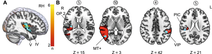
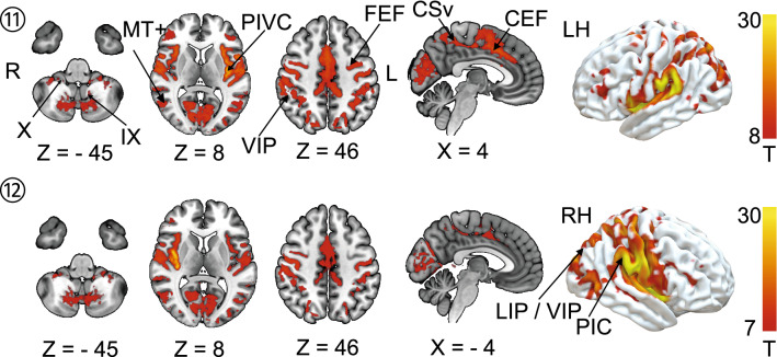
In rare cases, cortical infarcts lead to vertigo. We evaluated structural and functional disconnection in patients with acute vertigo due to unilateral ischemic cortical infarcts compared to infarcts without vertigo in a similar location with a focus on the connectivity of the vestibular cortex, i.e., the parieto-opercular (retro-)insular cortex (PIVC). Using lesion maps from the ten published case reports, we computed lesion-functional connectivity networks in a set of healthy individuals from the human connectome project. The probability of lesion disconnection was evaluated by white matter disconnectome mapping. In all ten cases with rotational vertigo, disconnections of interhemispheric connections via the corpus callosum were present but were spared in lesions of the PIVC without vertigo. Further, the arcuate fascicle was affected in 90% of the lesions that led to vertigo and spared in lesions that did not lead to vertigo. The lesion-functional connectivity network included vestibulo-cerebellar hubs, the vestibular nuclei, the PIVC, the retro-insular and posterior insular cortex, the multisensory vestibular ventral intraparietal area, motion-sensitive areas (temporal area MT+ and cingulate visual sulcus) as well as hubs for ocular motor control (lateral intraparietal area, cingulate and frontal eye fields). However, this was not sufficient to differentiate between lesions with and without vertigo. Disruption of interhemispheric connections of both PIVC via the corpus callosum and intra-hemispheric disconnection via the arcuate fascicle might be the distinguishing factor between vestibular cortical network lesions that manifest with vertigo compared to those without vertigo.

Keywords: Corpus callosum; Cortical vertigo; Disconnectome; Insula; PIVC; Vestibular.


Conflict of interest statement

The authors have not disclosed any competing interests.

Figures

Fig. 1
Fig. 1
A Representative axial slices for each lesion of the published case reports on hemispheric infarcts that led to vertigo (lesions 1–10) and infarcts that did not present with vertigo (lesion overlap of 10 cases; 11–12). Left-sided lesions with vertigo are shown in the top row. Lesions were manually delineated in MNI152 space using anatomical landmark neighboring relationships
Fig. 2
Fig. 2
A Overlap image of ischemic infarcts involving the insula (overlap of at least 2 lesions is depicted): the ischemic lesions were mainly located in the posterior parts of the insula (anterior long insular gyrus and posterior long insular gyrus—IV, V). B In rare cases, the lesions involved vestibular structures in the parietal operculum (OP2), the intraparietal sulcus (area VIP) and the temporal lobe (MT+). None of the lesions directly affected area PIC; however, lesion 5 is in close proximity to this structure. OP2 parietal operculum 2; MT+ , motion-sensitive middle temporal area; VIP ventral intraparietal area; PIC posterior insular cortex, IV anterior long insular gyrus, V posterior long insular gyrus
Fig. 3
Fig. 3
Disconnectome map of all ten lesions that manifested with vertigo. Disconnectome results are thresholded at 0.5 (> 50% probability of disconnection). Disconnection was observed for the posterior fronto-insular tracts (IV, V) that connect the posterior insula with the opercula. Intra-hemispheric disconnection affected the arcuate fascicle and the superior longitudinal fascicle (SLF, mostly third branch). Interhemispheric disconnection was observed in the splenium of the corpus callosum. Additionally, disconnection was observed with the vestibular nuclei group in the pontomedullary brainstem (possibly via the medial longitudinal fascicle and medial lemniscus) and also with the cerebellum via the superior cerebellar peduncle
Fig. 4
Fig. 4
Overlap of functional connectivity networks (FCN) of the individual lesions (positive correlations with the lesion site). The individual lesion network maps are thresholded at p < 0.001, FWE corrected for multiple comparisons after calculating 10,000 permutations using TFCE. The figure shows an overlap of eight or more cases. Common vestibular network hubs to all lesions include the cerebellar vestibular and ocular motor representations in lobules IX (nodulus, uvula), lobule X (flocculus/paraflocculus), the vestibular nuclei (VN), the thalamus, the parieto-insular vestibular cortex (PIVC; includes insular gyri IV, V (anterior and posterior long insular gyri), area OP2, and retro-insular cortex), the posterior insular cortex (area PIC), motion-sensitive temporal and cingulate areas MT + and CSv (cingulate visual area), ocular motor and vestibular intraparietal areas LIP and VIP (lateral and ventral intraparietal area), the frontal and cingulate eye fields (FEF, CEF). The color bar gives the number of overlapping lesion FCNs
Fig. 5
Fig. 5
Shows the FCN of the right- and left-sided lesions of the posterior insula that did not lead to vertigo/dizziness (n = 10 cases). The FCN observed in these lesions was remarkably similar, involving the vestibulo-cerebellum and the cortical vestibular and ocular motor networks. All results thresholded at p < 0.001, FWE corrected for multiple comparisons on the cluster level after 10 000 permutations using TFCE. The color bar shows t score intensities. IX, X cerebellar lobules IX, X, MT+ motion-sensitive temporal area, PIVC parieto-opercular (retro-)insular vestibular cortex, PIC posterior insular cortex, FEF frontal eye field, CEF cingulate eye field, LIP/VIP lateral/ventral intraparietal area, CsV cingulate visual sulcus

References

    1. Aboitiz F, Montiel J. One hundred million years of interhemispheric communication: the history of the corpus callosum. Braz J Med Biol Res. 2003;36:409–420. doi: 10.1590/s0100-879x2003000400002. - DOI - PubMed
    1. Ahn B-Y, Bae J-W, Kim D-H, et al. Pseudovestibular neuritis associated with isolated insular stroke. J Neurol. 2010;257:1570–1572. doi: 10.1007/s00415-010-5547-y. - DOI - PubMed
    1. Anagnostou E, Spengos K, Vassilopoulou S, et al. Incidence of rotational vertigo in supratentorial stroke: a prospective analysis of 112 consecutive patients. J Neurol Sci. 2010;290:33–36. doi: 10.1016/j.jns.2009.11.015. - DOI - PubMed
    1. Angelaki DE, Cullen KE. Vestibular system: the many facets of a multimodal sense. Annu Rev Neurosci. 2008;31:125–150. doi: 10.1146/annurev.neuro.31.060407.125555. - DOI - PubMed
    1. Avants BB, Tustison NJ, Song G, et al. A reproducible evaluation of ANTs similarity metric performance in brain image registration. Neuroimage. 2011;54:2033–2044. doi: 10.1016/j.neuroimage.2010.09.025. - DOI - PMC - PubMed

LinkOut - more resources