. 2022 Jul 15;13(1):117.
doi: 10.1186/s13244-022-01246-z.

The current role and future directions of imaging in failed back surgery syndrome patients: an educational review

Affiliations

The current role and future directions of imaging in failed back surgery syndrome patients: an educational review

Richard L Witkam et al. Insights Imaging. .

Abstract

Background: Failed back surgery syndrome (FBSS) is an umbrella term referring to painful sensations experienced by patients after spinal surgery, mostly of neuropathic nature. Adequate treatment of FBSS is challenging, as its etiology is believed to be multifactorial and still not fully clarified. Accurate identification of the source of pain is difficult but pivotal to establish the most appropriate treatment strategy. Although the clinical utility of imaging in FBSS patients is still contentious, objective parameters are highly warranted to map different phenotypes of FBSS and tailor each subsequent therapy.

Main body: Since technological developments have weakened the applicability of prior research, this educational review outlined the recent evidence (i.e., from January 2005 onwards) after a systematic literature search. The state of the art on multiple imaging modalities in FBSS patients was reviewed. Future directions related to functional MRI and the development of imaging biomarkers have also been discussed.

Conclusion: Besides the fact that more imaging studies correlated with symptomatology in the postoperative setting are warranted, the current educational review outlined that contrast-enhanced MRI and MR neurography have been suggested as valuable imaging protocols to assess alterations in the spine of FBSS patients. The use of imaging biomarkers to study correlations between imaging features and symptomatology might hold future potential; however, more research is required before any promising hypotheses can be drawn.

Keywords: Artificial intelligence; Diagnostic imaging; Failed back surgery syndrome; Magnetic resonance imaging; Signs and symptoms.


Conflict of interest statement

The authors declare that they have no competing interests.
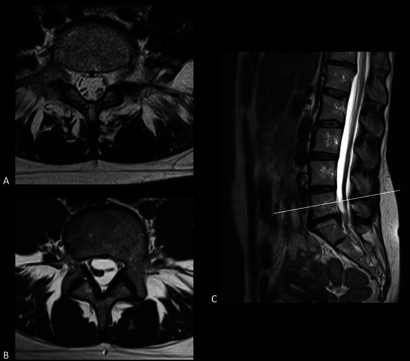
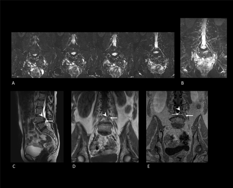
Figures

Fig. 1
Fig. 1
Search strategy. Overall, we noticed that many studies did not provide a conclusive statement on what patients were studied or what symptoms the studied patients suffered from. Consequently, these studies were excluded as we were unsure whether FBSS patients were studied. Also, some studies focused on the preoperative setting, and only a few studies explored any possible correlations between imaging features and symptomatology. Since the reviewed papers in the current study were nonrandomized and mostly explorative, no quality assessment tools were available. Therefore, the level of evidence and risk of bias could not formally be assessed. The lack of randomized or diagnostic accuracy trials could be declared by the current obscurity of what imaging features may clarify FBSS symptomatology
Fig. 2
Fig. 2
Radiographs of the thoracic- and lumbar spine and pelvic region of an FBSS patient. A The spinal cord stimulation electrode projects centrally at the level of Th8–Th10 on the anterior–posterior view. B The lateral view shows that the electrode projects over the posterior one-third of the spinal canal at levels Th8–Th10. C, D The internalized pulse generator can be observed projecting over the left gluteal region. Extension cables can be observed between the internalized pulse generator and the spinal cord stimulation electrode. Osteosynthesis materials can be observed projecting over the sacroiliac joint bilaterally and the lumbosacral joint. The vertebral column shows no anterior or posterior displacement. Degenerative changes in the thoracic and lumbar vertebrae can be observed with anterior and lateral osteophytes
Fig. 3
Fig. 3
Lumbar spine CT images in an FBSS patient with an implanted spinal cord stimulation system. AC Sagittal reconstructions through the vertebral column as depicted by the axial miniature on the right-hand side. The spinal cord stimulation electrode can be observed lining the ventral aspect of the dorsal lamina on level Th12–L1. Sclerosis of L5 and S1 can be observed. Additionally, at level L5–S1, bilateral laminectomy has been carried out. D Three-dimensional reconstructions of the spine of the same FBSS patient. The connection of the extension cables to the internalized pulse generator can be appreciated in blue at level L3–L4. E Virtually dissected three-dimensional model of the spine of the same FBSS patient. The spinal cord stimulation electrode can be appreciated as multiple blue dots in the spinal canal. The extension cable can be followed into the paraspinal space
Fig. 4
Fig. 4
Fluoroscopic myelography and CT myelography images after lumbar punction and intradural injection with iodine-contrast medium. A Fluoroscopic myelography image after lumbar punction. The lumbar punction needle is annotated by the white arrow. Iodine-contrast is inserted into intradural space and shows spinal stenosis (white arrowheads) at level L4–L5 and, to a lesser extent, at level L3–L4. BD Sagittal reconstructions of CT myelography (localization as depicted by the axial miniature on the right-hand side). The injected iodine-contrast is observed as radiopaque fluid in the dural sac. These images confirmed the observed spinal stenosis at level L4–L5 and L3–L4 as observed on fluoroscopic myelography. Spinal stenoses are annotated by use of the white arrowheads
Fig. 5
Fig. 5
Planar skeletal scintigraphy of an FBSS patient using technetium-99m hydroxydiphosphonate (Tc-99m HDP) to investigate rheumatological disease in the tarsal and metatarsal joints. A Posterior full-body planar image; B anterior full-body planar image; C Lateral skull planar image. Although Tc-99m HDP skeletal scintigraphy was carried out to assess the tarsal and metatarsal joints, full-body planar images were also acquired. In corroboration with the findings of Huang et al. [29], planar images show no specific features of FBSS. Findings were consistent with (nonspecific) degenerative changes in the spinal column and the bilateral tarsometatarsal joints. Focal round accumulation of tracer was observed in near the left parietal bone. This incidental finding was consistent with left parietal meningioma
Fig. 6
Fig. 6
SPECT/CT of an FBSS patient using Tc-99m HDP to investigate the cause of persistent pain after left-sided laminectomy at level L5–S1. Focal radiotracer accumulation was observed at level L4–L5, both near the intervertebral disk and the right zygapophyseal joint. Tracer accumulation was considered to reflect active osteoarthritic changes. No reoperation was carried out
Fig. 7
Fig. 7
MRI of the lumbar spine demonstrates a large disk herniation on level L5–S1. A Sagittal T2-weighted MRI depicts disk protrusion at level L5–S1. A close relation with the cauda equina roots can be observed. B Axial T2-weighted MRI shows the protruding disk at level L5–S1 occupying part of the central zone and right subarticular zone. Cauda equina roots are displaced to the contralateral side of the dural sac
Fig. 8
Fig. 8
Axial MRI of the postoperative lumbar spine at level L4–L5. A Axial T2-weighted image showing soft tissue occupying the subarticular zone bilaterally. Tissue shows to have an intermediate signal intensity. Cauda equina roots are displaced and course more central in the dural sac. B Axial T1-weighted image showing the tissue characteristics of the known soft tissue in the bilateral subarticular zone. On native T1-weighted images, an intermediate signal intensity is observed. C Post-contrast axial T1-weighted image showing homogeneous enhancement of the known soft tissue in the subarticular zone bilaterally. Based on this enhancement, a suggestion of postoperative epidural fibrosis was made
Fig. 9
Fig. 9
T2-weighted MRI of the same FBBS patient at two-time intervals. A First available axial T2-weighted MRI section at the level of the fifth lumbar vertebra. Cauda equina roots are seen to float in ample cerebrospinal fluid. B Axial T2-weighted MRI section at the level of the fifth lumbar vertebra of the same patient after spinal intervention at level L5–S1. The patient complains of recurrent right-sided leg pain, sensory changes, and motor weakness. MRI show new clumping of the cauda equina roots on the right side of the dural sac. These features were consistent with arachnoiditis. C Mid-sagittal T2-weighted MRI, which also shows the clumping of cauda equina roots
Fig. 10
Fig. 10
MR neurography examination of two FBBS patients. A Subsequent coronal 3D short tau inversion recovery sequence (3D STIR) images of one patient with no abnormalities. B Maximum-intensity projection of the 3D STIR images of the same patient (as depicted in A) showing no abnormalities of the lumbar plexus. C Sagittal T2-weighted MRI of a different patient showing a disk herniation occupying the left subarticular zone at level L4–L5 (white arrowhead). The white arrow depicts the coursing spinal nerve L4 on the left. D Coronal T1-weighted MRI of the same patient (as depicted in C, E) showing the disk herniation (white arrowhead) and left L4 spinal nerve (white arrow). E Coronal T1-weighted post-contrast MRI showing enhancing tissue (white dotted arrow) surrounding the disk herniation (white arrowhead). In addition, subtle perineural enhancement of the left L4 spinal nerve (white arrow) can be appreciated

Similar articles

Cited by

References

    1. Schug SA, Lavand'homme P, Barke A, et al. The IASP classification of chronic pain for ICD-11: chronic postsurgical or posttraumatic pain. Pain. 2019;160(1):45–52. doi: 10.1097/j.pain.0000000000001413. - DOI - PubMed
    1. Christelis N, Simpson B, Russo M, et al. Persistent spinal pain syndrome: a proposal for failed back surgery syndrome and ICD-11. Pain Med. 2021;22(4):807–818. doi: 10.1093/pm/pnab015. - DOI - PMC - PubMed
    1. Merskey H, Bogduk N. Classification of chronic pain: descriptions of chronic pain syndromes and definitions of pain terms. 2. IASP Press; 1994. p. 222.
    1. Manca A, Eldabe S, Buchser E, et al. Relationship between health-related quality of life, pain, and functional disability in neuropathic pain patients with failed back surgery syndrome. Value Health. 2010;13(1):95–102. doi: 10.1111/j.1524-4733.2009.00588.x. - DOI - PubMed
    1. Thomson S, Jacques L. Demographic characteristics of patients with severe neuropathic pain secondary to failed back surgery syndrome. Pain Pract. 2009;9(3):206–215. doi: 10.1111/j.1533-2500.2009.00276.x. - DOI - PubMed

LinkOut - more resources