. 2022 Sep:174:92-100.
doi: 10.1016/j.radonc.2022.07.008. Epub 2022 Jul 15.

Targeting HER3-dependent activation of nuclear AKT improves radiotherapy of non-small cell lung cancer

Affiliations

Targeting HER3-dependent activation of nuclear AKT improves radiotherapy of non-small cell lung cancer

Mahmoud Toulany et al. Radiother Oncol. 2022 Sep.

Abstract

Background: AKT1 must be present and activated in the nucleus immediately after irradiation to stimulate AKT1-dependent double-strand breaks (DSB) repair through the fast non-homologous end-joining (NHEJ) repair process. We investigated the subcellular distribution of AKT1 and the role of HER family receptor members on the phosphorylation of nuclear AKT and radiation response.

Materials and methods: Using genetic approaches and pharmacological inhibitors, we investigated the subcellular distribution of AKT1 and the role of HER family receptor members on the activation of nuclear AKT in non-small cell lung cancer (NSCLC) cells in vitro. ɤH2AX foci assay was applied to investigate the role of AKT activating signaling pathway on DSB repair. A mouse tumor xenograft model was used to study the impact of discovered signaling pathway activating nuclear AKT on the radiation response of tumors in vivo.

Results: Our data suggests that neither ionizing radiation (IR) nor stimulation with HER family receptor ligands induced rapid nuclear translocation of endogenous AKT1. GFP-tagged exogenous AKT1 translocated to the nucleus under un-irradiated conditions and IR did not stimulate this translocation. Nuclear translocation of GFP-AKT1 was impaired by the AKT inhibitor MK2206 as shown by its accumulation in the cytoplasmic fraction. IR-induced phosphorylation of nuclear AKT was primarily dependent on HER3 expression and tyrosine kinase activation of epidermal growth factor receptor. In line with the role of AKT1 in DSB repair, the HER3 neutralizing antibody patritumab as well as HER3-siRNA diminished DSB repair in vitro. Combination of patritumab with radiotherapy improved the effect of radiotherapy on tumor growth delay in a xenograft model.

Conclusion: IR-induced activation of nuclear AKT occurs inside the nucleus that is mainly dependent on HER3 expression in NSCLC. These findings suggest that targeting HER3 in combination with radiotherapy may provide a logical treatment option for investigation in selected NSCLC patients.

Keywords: DNA repair; HER3; Non-small cell lung cancer; Nuclear AKT; Radioresistance.


Conflict of interest statement

Conflicts of interest

All authors declare no competing financial interests.

Figures

Fig. 1
Fig. 1
IR does not induce AKT1 nuclear translocation. Cells were serum starved for 24 hours and irradiated 4 Gy. Cytoplasmic and nuclear fractions were isolated as described [18] at indicates time-points after irradiation and subjected tom SDS-PAGE. Expression level of AKT1 was analyzed by Western blotting. α-Tubulin and H3 were detected as cytoplasmic and nuclear markers, respectively.
Fig. 2
Fig. 2
AKT inhibition blocks nuclear translocation of AKT1. (A) A549 cells were transfected with plasmids expressing GFP or GFP-AKT1 and followed by treatment with AKT inhibitor MK2206 (10 μM) either 24 h after transfection (A) or simultaneously at the time of transfection (C). The cytoplasmic and nuclear fractions were isolated 24 h after treatment with the inhibitor. (A, C) Level of p-AKT (S473), AKT1, GFP were detected by Western blotting. α-Tubulin and lamin A/C were detected as cytoplasmic and nuclear markers, respectively. The data shown are representative of three independent experiments. (B) Mean density of GFP-AKT1 from 3 independent experiments. The asterisk indicates a significant increase in cytoplasmic GFP-AKT1 after treatment with MK2206 (10 μM) compared to that in DMSO treated cells (* p < 0.05, students t -test).
Fig.3
Fig.3
Stimulation with HER3- but not EGFR ligand enhances AKT phosphorylation in the nucleus. A549 cells were serum starved for 24 hours (A) or cultured in medium containing 10% FCS (B) and treated with TGFα (100 ng/ml) (A) or NRG (50 ng/ml) (B) for indicated time-points. Thereafter, cytoplasmic and nuclear fractions were isolated and subjected to SDS-PAGE. Indicated phospho- and total proteins were detected by Western blotting. Phospho-AKT-S473, AKT1 and HER3 were detected from the first blot. (B) EGFR, P-HER3, Phospho-AKT-T308 and the cytoplasmic/nuclear markers (Tubulin and lamin A/C) were detected from the second blot. (A&B) Phospho-AKT and phospho-EGFR blots were stripped and incubated with antibodies against corresponding total protein. α-Tubulin and Lamin A/C were detected as cytoplasmic and nuclear markers, respectively. Densitometry values are normalized to the control non-stimulated condition in nuclear fraction.
Fig. 4
Fig. 4
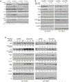
IR-induced phosphorylation of nuclear AKT is HER2- and HER3- but not EGFR-dependent. A549 cells were reverse transfected with 10 nM of indicated siRNA and 48 h after transfection were serum starved. Twenty-four hours later, cells were irradiated with 4 Gy (A–B) or stimulated with NRG (10 nM) (B). (C) A549 ells were transfected with indicated plasmids and incubated for 24 h. Thereafter, cells were starved for additional 24 h and followed by irradiation. At the indicated time-points after irradiation (A–C) and 10 min after NRG treatment (B) total lysates (A) and sub-cellular protein factions (B, C) were isolated and subjected to SDS-PAGE. Thereafter, level of activation and expression of indicated proteins was analyzed by Western blotting. α-Tubulin and H3 were detected as cytoplasmic and nuclear markers, respectively. MW: Molecular Weight.
Fig. 5
Fig. 5
Targeting HER3 blocks activation of nuclear AKT, impairs DSB repair and improve radiotherapy outcome. A549 cells were treated with indicated antibodies at the concentrations described in the results section for one hour and mock irradiated or irradiated with 4 Gy. Total protein lysates (A) and subcellular fractions (B) were isolated at the indicated time-points after irradiation. The activity and the expression profile of the indicated proteins were analyzed by Western blotting. α-Tubulin and H3 were detected as cytoplasmic and nuclear markers, respectively. (C–D) Cells were reverse transfected with 10 nM of indicated siRNA (C) or treated with patritumab (100 μg/ml, for 24 h) (D) and irradiated 48 h after transfection or 24 h after treatment with patritumab. γH2AX foci assay was performed 24 h after irradiation. Mean number of foci was calculated in nuclei of at least 340 cells from 2 independent experiments and graphed. (E) Nude female mice were injected with A549 (2 × 10⁶ cells) subcutaneously and tumor diameters were measured 2 times per week until the end of the experiment. Upon reaching tumors to approximately 400 mm3, animals were treated with either IgG or patritumab (4 mg/kg) and followed with or without irradiation, as described in Materials and Methods section. Data presents mean tumor volume ± SEM of 10 tumors (5 mice). Asterisks indicate a significant difference in number of residual DSB (*** p < 0.001, students t -test) (C–D) and tumor growth (E) between the indicated groups (** p < 0.01, nonparametric Friedman test). MW: Molecular Weight.

Similar articles

Cited by

References

    1. Knighton DR, Cadena DL, Zheng J, Ten Eyck LF, Taylor SS, Sowadski JM, et al. Structural features that specify tyrosine kinase activity deduced from homology modeling of the epidermal growth factor receptor. Proc Natl Acad Sci U S A 1993;90:5001–5005. doi:10.1073/pnas.90.11.5001. - DOI - PMC - PubMed
    1. Black L, Longo JF, Carroll SL Mechanisms of ERBB3 action in human neoplasia. Am J Pathol 2019;189:1898–1912. doi:10.1016/j.ajpath.2019.06.008. - DOI - PMC - PubMed
    1. Mujoo K, Choi B-K, Huang Z, Zhang N, An Z Regulation of ERBB3/HER3 signaling in cancer. Oncotarget 2014;5:10222–10236. doi:10.18632/oncotarget.2655. - DOI - PMC - PubMed
    1. Oh D-Y, Bang Y-J HER2-targeted therapies – a role beyond breast cancer. Nat Rev Clin Oncol 2020;17:33–48. doi:10.1038/s41571-019-0268-3. - DOI - PubMed
    1. Meric-Bernstam F, Johnson AM, Dumbrava EEI, Raghav K, Balaji K, Bhatt M, et al. Advances in HER2-targeted therapy: novel agents and opportunities beyond breast and gastric cancer. Clin Cancer Res 2019;25:2033–2041. doi:10.1158/1078-0432.CCR-18-2275. - DOI - PMC - PubMed

Publication types