. 2022 Aug 10;42(32):6195-6210.
doi: 10.1523/JNEUROSCI.1395-21.2022. Epub 2022 Jul 15.

The MAP3Ks DLK and LZK Direct Diverse Responses to Axon Damage in Zebrafish Peripheral Neurons

Affiliations

The MAP3Ks DLK and LZK Direct Diverse Responses to Axon Damage in Zebrafish Peripheral Neurons

Kadidia Pemba Adula et al. J Neurosci. .

Abstract

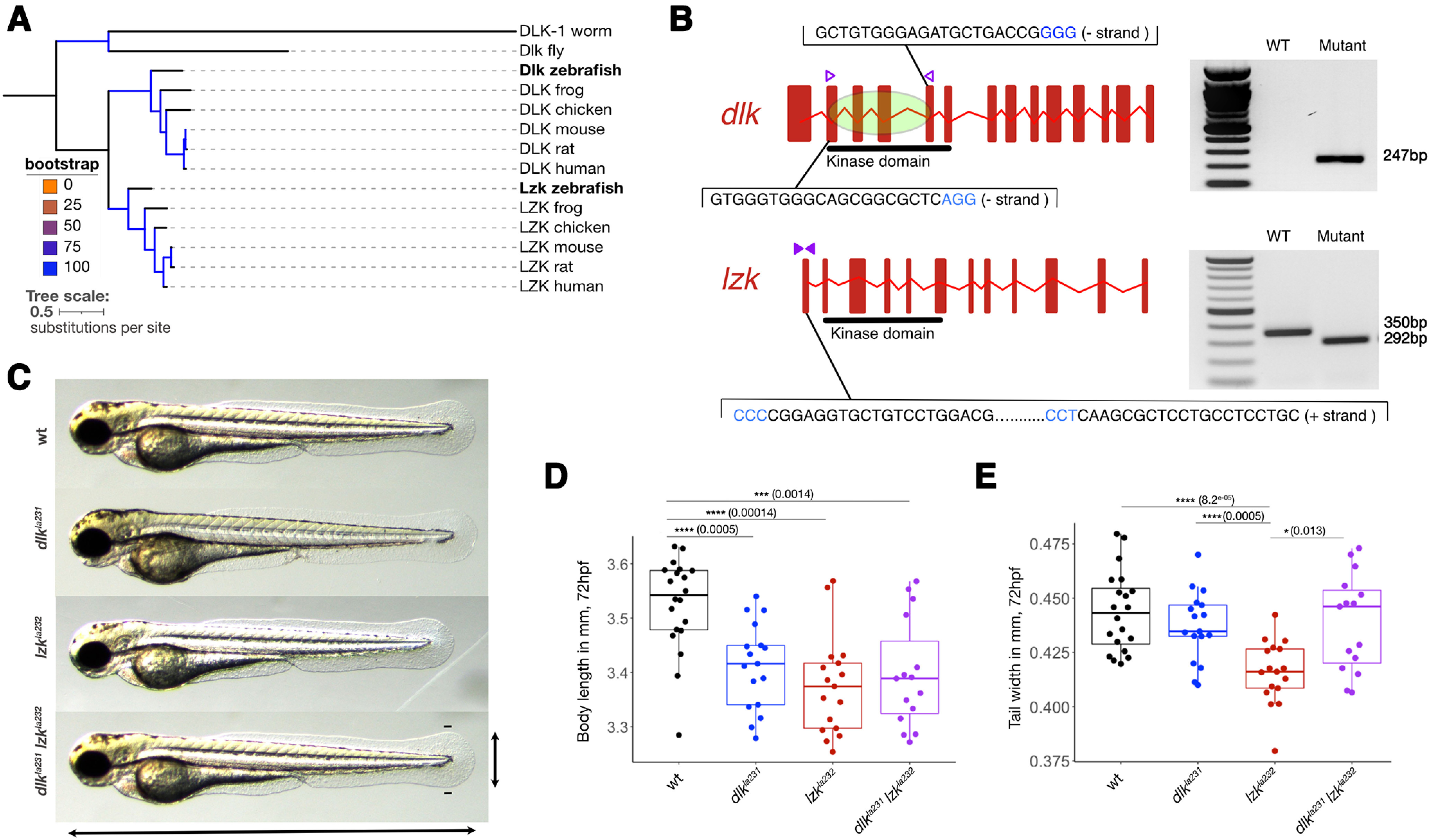
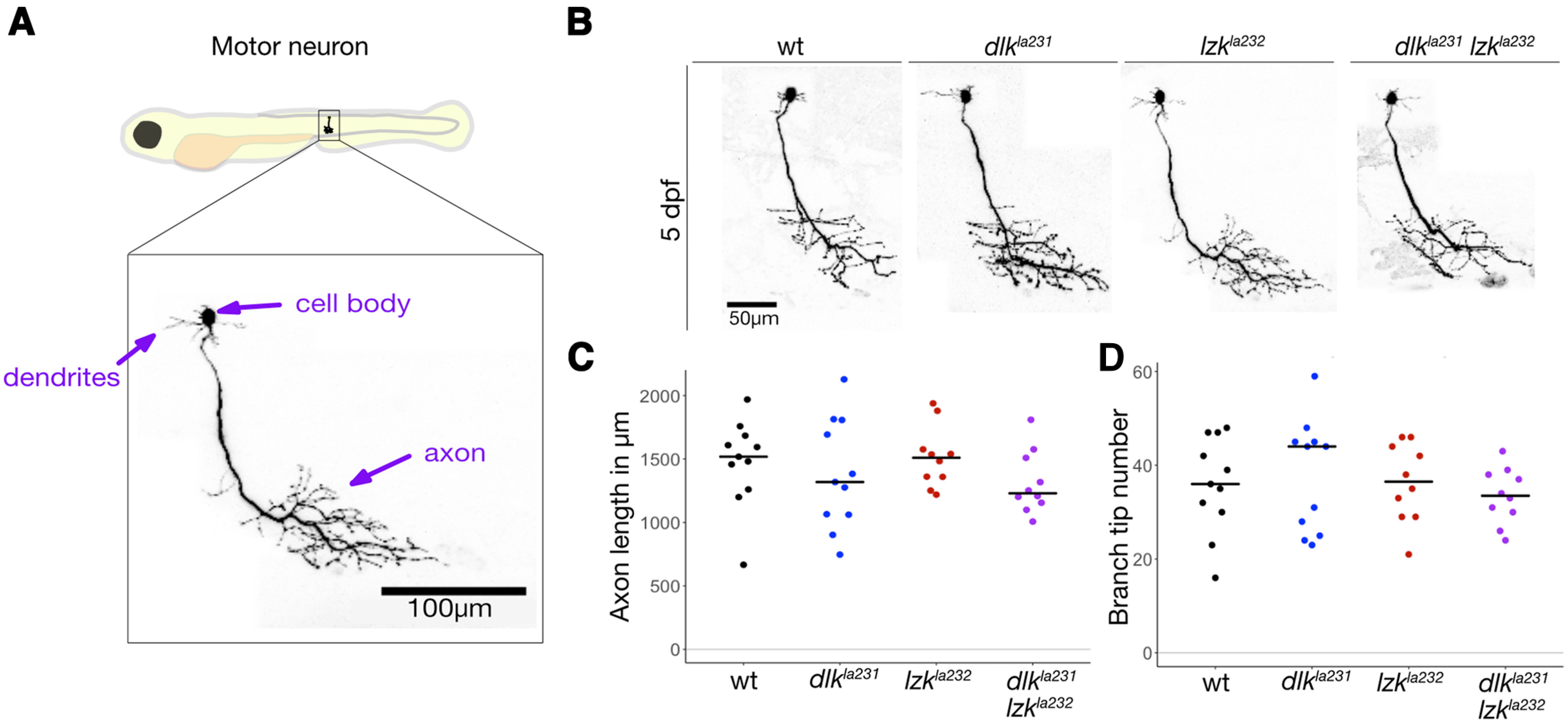
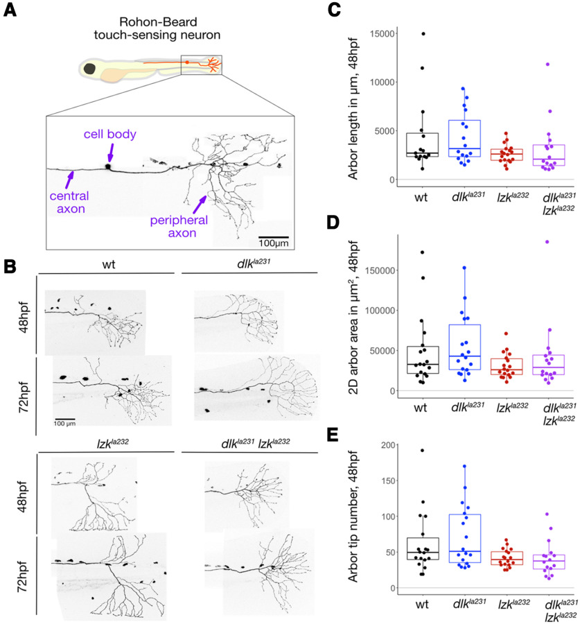
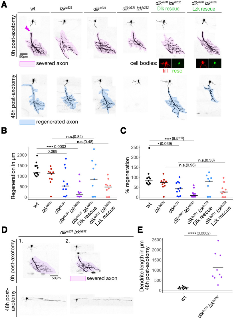
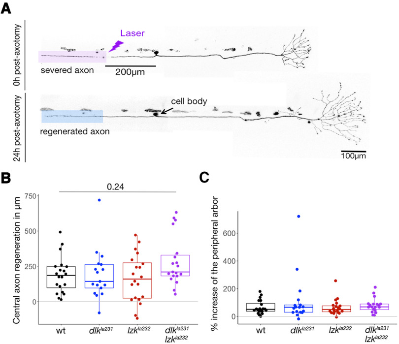
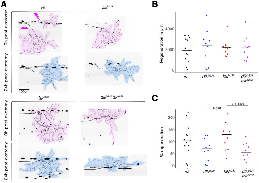
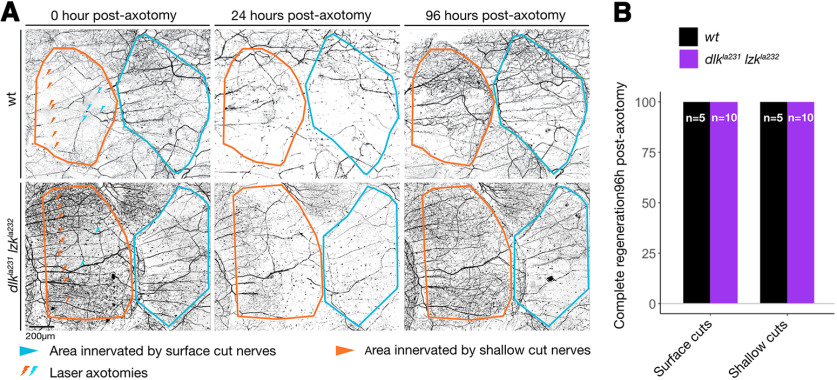
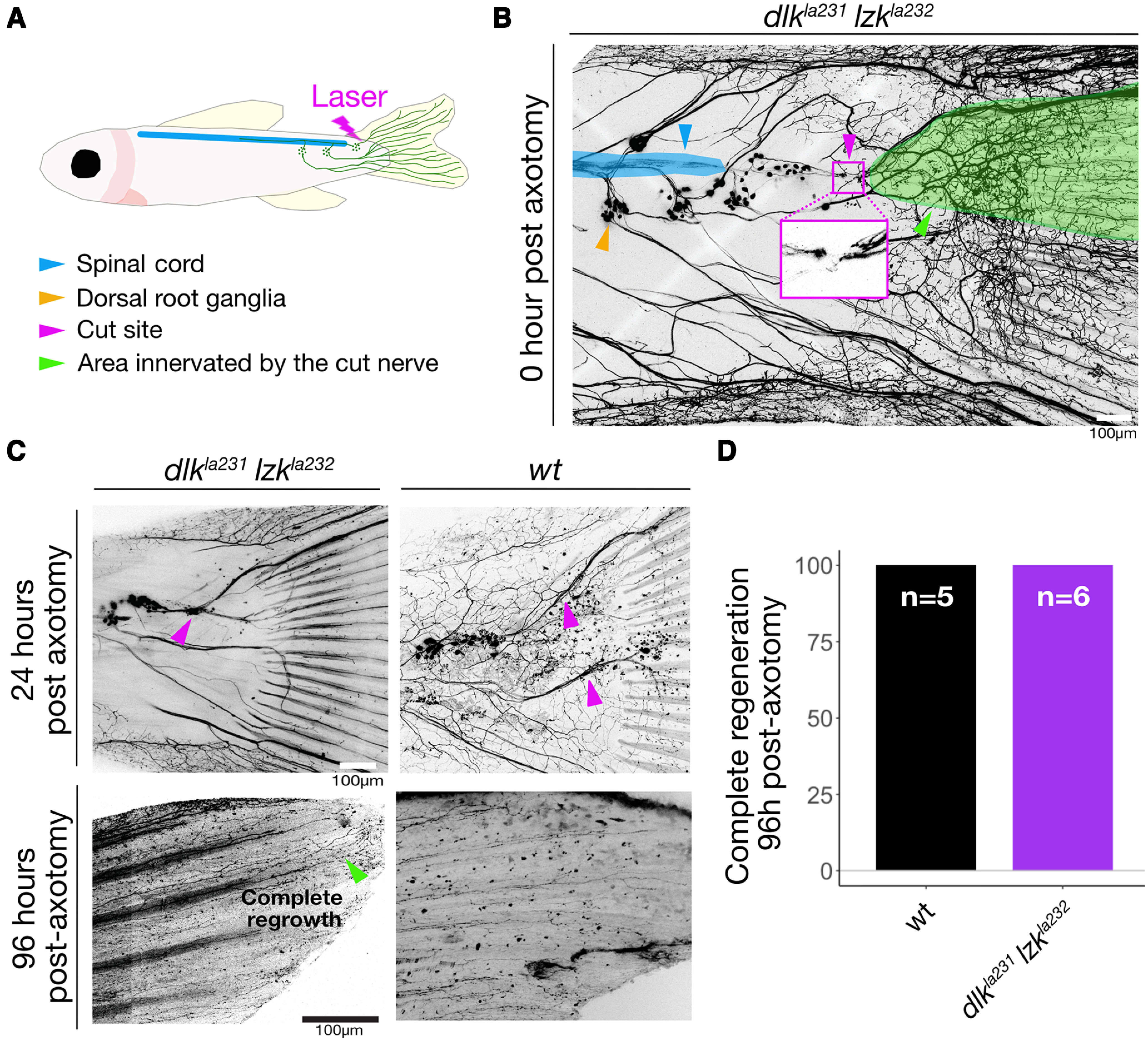
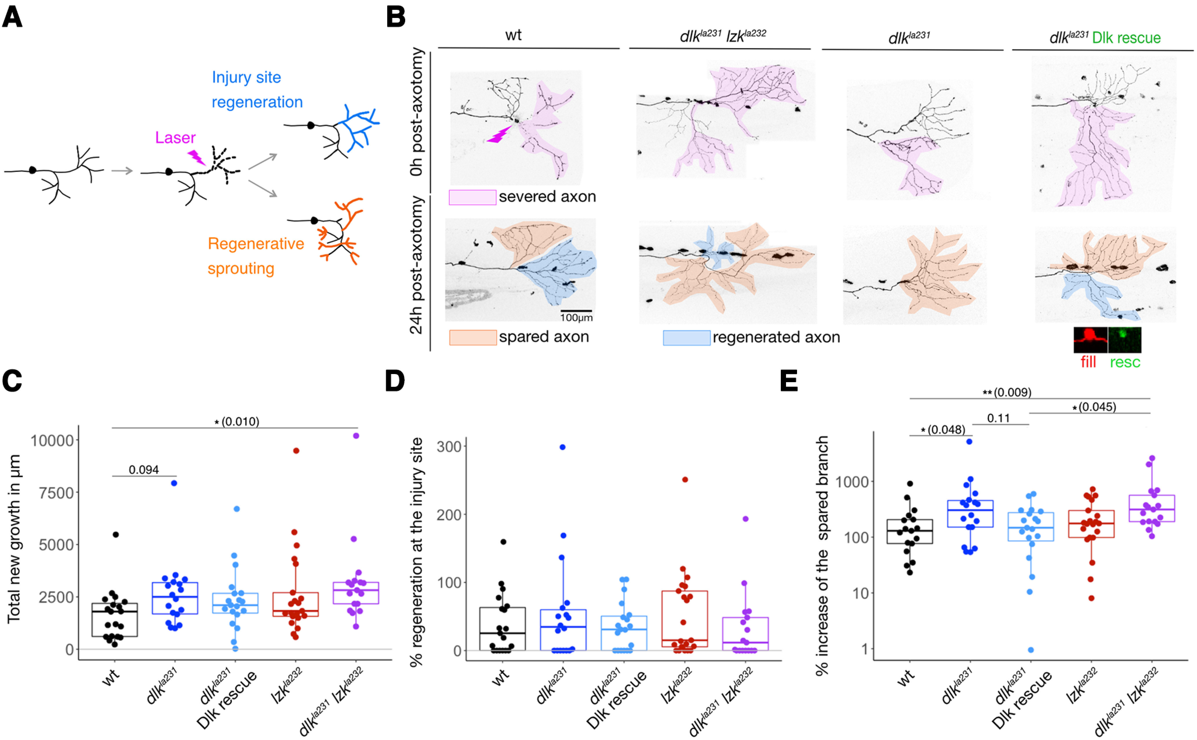
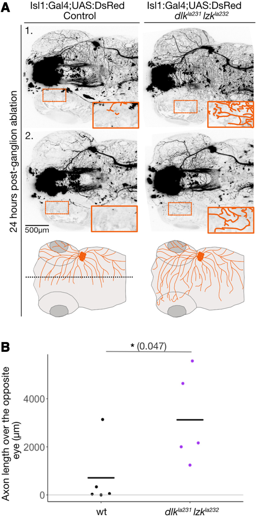
Mitogen-activated protein kinase kinase kinases (MAP3Ks) dual leucine kinase (DLK) and leucine zipper kinase (LZK) are essential mediators of axon damage responses, but their responses are varied, complex, and incompletely understood. To characterize their functions in axon injury, we generated zebrafish mutants of each gene, labeled motor neurons (MNs) and touch-sensing neurons in live zebrafish, precisely cut their axons with a laser, and assessed the ability of mutant axons to regenerate in larvae, before sex is apparent in zebrafish. DLK and LZK were required redundantly and cell autonomously for axon regeneration in MNs but not in larval Rohon-Beard (RB) or adult dorsal root ganglion (DRG) sensory neurons. Surprisingly, in dlk lzk double mutants, the spared branches of wounded RB axons grew excessively, suggesting that these kinases inhibit regenerative sprouting in damaged axons. Uninjured trigeminal sensory axons also grew excessively in mutants when neighboring neurons were ablated, indicating that these MAP3Ks are general inhibitors of sensory axon growth. These results demonstrate that zebrafish DLK and LZK promote diverse injury responses, depending on the neuronal cell identity and type of axonal injury.SIGNIFICANCE STATEMENT The MAP3Ks DLK and LZK are damage sensors that promote diverse outcomes to neuronal injury, including axon regeneration. Understanding their context-specific functions is a prerequisite to considering these kinases as therapeutic targets. To investigate DLK and LZK cell-type-specific functions, we created zebrafish mutants in each gene. Using mosaic cell labeling and precise laser injury we found that both proteins were required for axon regeneration in motor neurons but, unexpectedly, were not required for axon regeneration in Rohon-Beard or DRG sensory neurons and negatively regulated sprouting in the spared axons of touch-sensing neurons. These findings emphasize that animals have evolved distinct mechanisms to regulate injury site regeneration and collateral sprouting, and identify differential roles for DLK and LZK in these processes.

Keywords: DLK; LZK; axon; regeneration; sprouting; zebrafish.


Figures

Figure 1.
Figure 1.
dlk And lzk zebrafish mutants. A, Phylogenetic tree of DLK and LZK orthologs. B, Genotyping of dlkla231 and lzkla232 CRISPR/Cas9 mutants. Left, gene structure of zebrafish dlk and lzk, with gRNA sequences. Blue indicates PAM (protospacer adjacent motif) sites. Right, DNA gel showing WT and mutant genotyping with primers indicated to the left (arrowheads). C, Forty-eight hpf zebrafish larvae of the indicated phenotypes. D, Overlaid box and dot plots comparing animal lengths from the tip of the head to the end of the tail each genotype. E, Overlaid box and dot plots comparing tail width in each genotype (see above, Materials and Methods; Table 5 for details of statistical analyses).
Figure 2.
Figure 2.
Motor neurons develop normally in dlkla231 and lzkla232 mutants. A, Diagram of 5 dpf larva, showing the approximate location of the image below of a single labeled MN in a live animal. The cell body and dendrites are in the spinal cord; the axon exits the spinal cord to innervate the ventral muscles of one segment. B, Labeled MNs in each of the indicated genotypes. C, Dot plot showing lengths of MNs in each of the indicated genotypes. Bar indicates the mean. There was no significant difference between groups (because distributions were normal, groups were compared by ANOVA). D, Dot plot showing branch tip numbers of MNs in each of the indicated genotypes. Bar indicates the mean. There was no significant difference between groups (see above, Materials and Methods; Table 5 for details of statistical analyses). Scale bars: A, 100 μm; B, 50 μm.
Figure 3.
Figure 3.
Rohon–Beard neurons develop normally in dlkla231 and lzkla232 mutants. A, Diagram of 48 hpf larva, showing the approximate location of the image below of a single labeled RB neuron in a live animal. The cell body and central and peripheral axons are labeled. The cell body and central axon are in the spinal cord; the peripheral axon exits the spinal cord to arborize in the developing epidermis. B, Tail-innervating peripheral RB axon arbors of the indicated genotypes at 48 and 72 hpf. C–E, Quantification of RB peripheral axon arbor length (C), 2D arbor area (D), and branch tip number (E) at 48 hpf (see above, Materials and Methods; Table 5 for details of statistical analyses). Scale bars: 100 μm.
Figure 4.
Figure 4.
Motor neuron regeneration is impaired in dlkla231 lzkla232 mutants. A, Top, Five days postfertilization motor axons immediately after axotomy in the indicated genotypes. Lightning bolt indicates axotomy site. Magenta highlights the separated distal stump that will degenerate. Far right, Neurons expressing rescue cDNAs; expression of rescue transgenes in cell bodies is shown below. Bottom, Same neurons 48 h postaxotomy. Blue highlights regenerated axons. B, C, Dot plots showing total regenerated length in each genotype (B) and the percentage of the original axon length regenerated (C). Bar indicates the mean. D, Dendrite overgrowth phenotype following failure of axon regeneration in dlkla231 lzkla232 mutants. E, Quantification of dendrite overgrowth (see above, Materials and Methods; Table 5 for details of statistical analyses). Scale bars: 50 μm.
Figure 5.
Figure 5.
RB central axons regenerate in dlkla231 lzkla232 mutants. A, Top, Forty-eight hours postfertilization RB axon immediately after axotomy. Magenta highlights the separated distal stump that will degenerate. Bottom, Same neuron 24 h postaxotomy. Blue highlights the regenerated axon. B, Overlaid box and dot plots showing central axon length regenerated in the indicated genotypes. C, Overlaid box and dot plots showing growth of peripheral arbors following central axotomy in the indicated genotypes (see above, Materials and Methods; Table 5 for details of statistical analyses). Scale bar, 100 μm.
Figure 6.
Figure 6.
RB peripheral axon arbors regenerate in dlkla231 lzkla232 mutants. A, Top, Forty-eight hours postfertilization RB peripheral axon immediately after axotomy of the indicated genotypes. Magenta highlights the separated distal stump that will degenerate. Bottom, Same neurons 24 h postaxotomy. Blue highlights the regenerated axon. B, C, Dot plots showing total regenerated length in each genotype (B) and the percentage of the original axon length regenerated (C). Bars indicate the mean (see above, Materials and Methods; Table 5 for details of statistical analyses). Scale bar, 100 μm.
Figure 7.
Figure 7.
DRG axons innervating the adult scale regenerate in dlkla231 lzkla232 mutants. A, Images of DRG neurons innervating the epidermis above scales in adult (8–11 months old) zebrafish in indicated genotypes at 0 h postaxotomy. Top, Immediately after axotomy; bottom, immediately before axotomy. Lightning bolts indicate axotomy sites of individual nerves growing into scales. Nerves innervating anterior scales (orange) are above the scale, whereas nerves innervating posterior scale (blue) are below the anterior scale. Most axons innervating these scales degenerated by 24 hpa (middle column) and reinnervated scales by 96 hpa (right columns). Scales were reinnervated in all wt (n = 10), dlkla231 lzkla232 (n = 5), dlkla231(n = 7, data not shown), and lzkla232 (n = 5, data not shown) mutants. B, Quantification of wt (n = 10) and dlkla231 lzkla232 (n = 5) mutant regeneration 96 hpa (see above, Materials and Methods; Table 5 for details of statistical analyses). Scale bar: 200 μm.
Figure 8.
Figure 8.
DRG axons innervating the juvenile tail regenerate in wild-type and dlkla231 lzkla232 mutants. A, Top left, Diagram of juvenile casper fish, showing tail-innervating DRGs in P2rx3a: LexA; 4xLexAop:mCherryla207transgenic fish. Lightning bolt indicates axotomy site. Blue arrow indicates the spinal cord. Legend refers to the image on the right. B, Overview showing the position of DRG cell bodies, sensory nerve layout, and an example of axotomy location in a 4- to 5-week-old dlkla231 lzkla232 mutant zebrafish. Inset, Axotomy site of the caudal-most DRG peripheral nerve. Green highlights separated arbors of the severed DRG nerve, which will degenerate after axotomy. C, Top left, Mutant dlkla231 lzkla232 showing regrowth of the severed nerve 24 h postaxotomy. Bottom left: Tail fin of the same animal showing that the regenerating axons have reached the fin tip. C, Top right, The wt fish showing regrowth of the severed nerve 24 hpa. Bottom right, Tail fin of the same animal showing that the regenerating axons have reached the fin tip. D, Quantification of complete regeneration at 96 h postaxotomy. Axons regenerated in all wt (n = 5) and all dlkla231 lzkla232 mutants (n = 6; see above, Materials and Methods; Table 5 for details of statistical analyses). Scale bars: 100 μm.
Figure 9.
Figure 9.
Spared arbors of damaged RB neurons sprout excessively in dlkla231 lzkla232 mutants. A, Diagram of partial RB peripheral axotomy assay, which differentiates between regeneration from the cut site and regenerative sprouting from spared branches. B, Top, Forty-eight hours postfertilization RB axons immediately after axotomy in the indicated genotypes. Lightning bolt indicates axotomy site. Magenta highlights the separated distal stump that will degenerate. Far right, A neuron expressing rescue cDNA; expression of the rescue transgene in the cell body is shown below. Bottom, Same neurons 24 h postaxotomy. Blue highlights regenerated axons; orange highlights the spared branch. C–E, Box and dot plots showing total new growth, including both from the axotomy site and spared branch (C), percentage regeneration from just the injury site (D), and percentage increase of the spared branch (E; see above, Materials and Methods; Table 5 for details of statistical analyses). Scale bar: 100 μm.
Figure 10.
Figure 10.
Trigeminal axons grow excessively in dlkla231 lzkla232 mutants after ablation of the contralateral ganglion. A, Images and diagram of trigeminal axons in zebrafish heads at 78 hpf. Confocal images show two separate examples of wt (left) and dlkla231 lzkla232 mutant (right) fish. Insets, Magnification of a region over the eye, with axons traced in orange. Bottom, Diagram depictions of the result. B, Dot plot showing total axon length that grew over the contralateral eye. Error bar indicates the mean (see above, Materials and Methods; Table 5 for details of statistical analyses). Scale bar, 500 μm.

Similar articles

Cited by

References

    1. Beckers A, Moons L (2019) Dendritic shrinkage after injury: a cellular killer or a necessity for axonal regeneration? Neural Regen Res 14:1313–1316. 10.4103/1673-5374.253505 - DOI - PMC - PubMed
    1. Beckers A, Van Dyck A, Bollaerts I, Van Houcke J, Lefevere E, Andries L, Agostinone J, Van Hove I, Di Polo A, Lemmens K, Moons L (2019) An antagonistic axon-dendrite interplay enables efficient neuronal repair in the adult zebrafish central nervous system. Mol Neurobiol 56:3175–3192. 10.1007/s12035-018-1292-5 - DOI - PubMed
    1. Chen L, Nye DM, Stone MC, Weiner AT, Gheres KW, Xiong X, Collins CA, Rolls MM (2016) Mitochondria and caspases tune Nmnat-mediated stabilization to promote axon regeneration. PLoS Genet 12:e1006503. 10.1371/journal.pgen.1006503 - DOI - PMC - PubMed
    1. Chen M, Geoffroy CG, Wong HN, Tress O, Nguyen MT, Holzman LB, Jin Y, Zheng B (2016) Leucine zipper-bearing kinase promotes axon growth in mammalian central nervous system neurons. Sci Rep 6:31482. 10.1038/srep31482 - DOI - PMC - PubMed
    1. Chen M, Geoffroy CG, Meves JM, Narang A, Li Y, Nguyen MT, Khai VS, Kong X, Steinke CL, Carolino KI, Elzière L, Goldberg MP, Jin Y, Zheng B (2018) Leucine zipper-bearing kinase is a critical regulator of astrocyte reactivity in the adult mammalian CNS. Cell Rep 22:3587–3597. 10.1016/j.celrep.2018.02.102 - DOI - PMC - PubMed

Publication types