Review
. 2022 Aug;43(8):630-639.
doi: 10.1016/j.it.2022.06.003. Epub 2022 Jul 12.

The potential role of HIV-1 latency in promoting neuroinflammation and HIV-1-associated neurocognitive disorder

Affiliations
Review

The potential role of HIV-1 latency in promoting neuroinflammation and HIV-1-associated neurocognitive disorder

Sheetal Sreeram et al. Trends Immunol. 2022 Aug.

Abstract

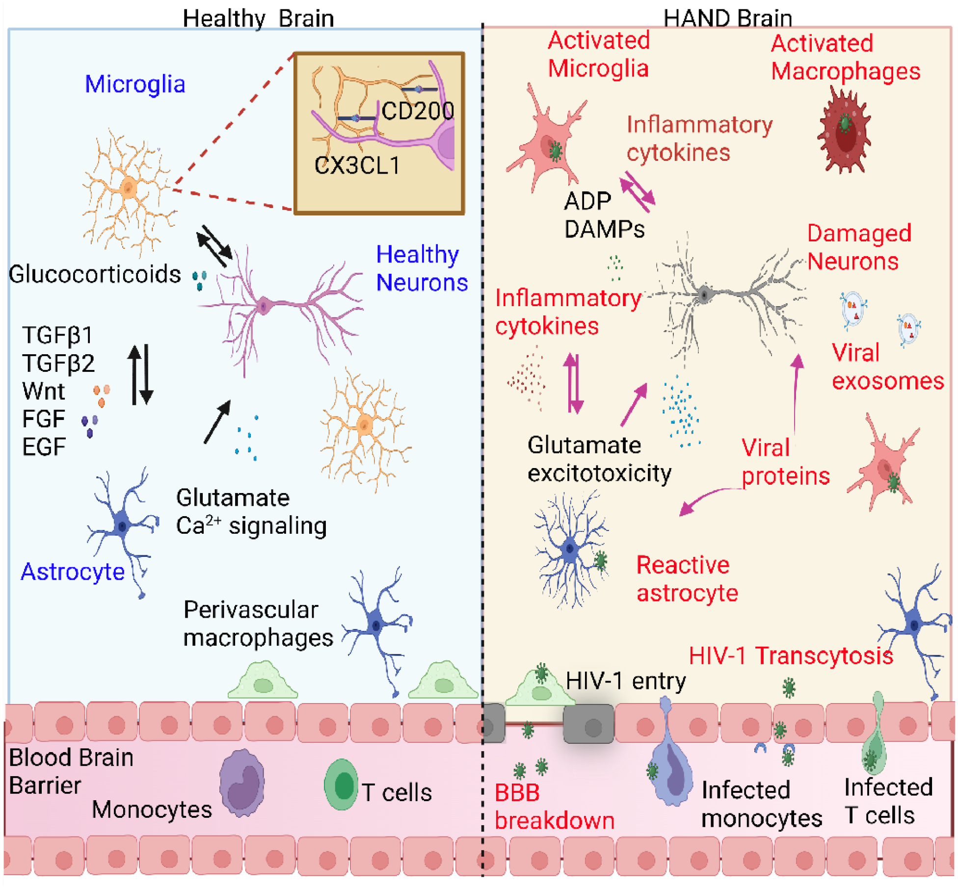
Despite potent suppression of HIV-1 viral replication in the central nervous system (CNS) by antiretroviral therapy (ART), between 15% and 60% of HIV-1-infected patients receiving ART exhibit neuroinflammation and symptoms of HIV-1-associated neurocognitive disorder (HAND) - a significant unmet challenge. We propose that the emergence of HIV-1 from latency in microglia underlies both neuroinflammation in the CNS and the progression of HAND. Recent molecular studies of cellular silencing mechanisms of HIV-1 in microglia show that HIV-1 latency can be reversed both by proinflammatory cytokines and by signals from damaged neurons, potentially creating intermittent cycles of HIV-1 reactivation and silencing in the brain. We posit that anti-inflammatory agents that also block HIV-1 reactivation, such as nuclear receptor agonists, might provide new putative therapeutic avenues for the treatment of HAND.

Keywords: HIV-1 latency; HIV-1-associated neurocognitive disorder; anti-inflammatory strategies; antiretroviral therapy; microglial cell activation.


Conflict of interest statement

Declaration of interests No interests are declared.

Figures

Figure 1.
Figure 1.. Schematic illustration of the interactions between neurons, astrocytes, and microglia in heathy vs. HAND human brains.
In a healthy brain, neurons and astrocytes induce microglia to attain a surveillance and neuroprotective phenotype by signaling though factors such as fractalkine CX3CL1, CD200, TGFβ1/2, and Wnt [77]. During HIV-1 infections, the “high-jacked” infected monocytes and CD4+ T cells allow viral entry into the brain. Viral particles can also enter the brain by damaging the blood-brain barrier (BBB), altering endothelial cell permeability, or through transcytosis [18]. HIV-1 can establish latent reservoirs in microglia. Reactivation of latent HIV-1 causes a microglial shift to a reactive state, resulting in the release of pro-inflammatory cytokines such as TNF-α, IL-1β, IL-6, IL-8, CCL2, and CCL5, reactive oxygen species (ROS), and reactive nitrogen species (RNS) [34]. Microglia activation can also cause astrocytes to shift to a reactive state, with secretion of more inflammatory cytokines [72]. Viral proteins such as gp120, Nef, Tat can directly cause neuronal damage and disrupt astrocyte glutamate transporters, resulting in glutamate imbalance in the brain and excitotoxicity of neurons [18,83]. Neural damage-associated molecular pattern (DAMP) molecules and ADP can further cause microglia hyperactivation and HIV-1 reactivation, thus initiating a cycle of progressive neural inflammation. This figure was created using BioRender.com.
Key Figure, Figure 2.
Key Figure, Figure 2.. Model for the regulation of HIV-1 transcription by inflammation and neuronal interactions.
HIV-1 can infect homeostatic (“ramified”) microglia. However, HIV-1 transcription might become quickly silenced by the recruitment of Nurr1 and the CoREST repressor complex to the long-term repeat regions, based on preliminary findings [48]. In the presence of “Off signals” produced by healthy neurons, HIV-1 infected cells are further suppressed by glucocorticoids (GC) [46] or other anti-inflammatory cytokines such as IL-4, IL-10, and TGF-β, produced by neurons. Latently infected cells can become fully reactive in the presence of exogenous pro-inflammatory activators, including LPS, GM-CSF, TNF-α and IFN-γ [33]. This leads to the activation of NF-κB, which can bind the LTR and activate HIV-1 transcription, releasing viral proteins such as Tat, Env, and Nef -- known neurotoxins. In addition to stimulating HIV-1 expression, NF-κB can activate a variety of cellular inflammatory markers and release additional neurotoxic cytokines including TNF-α, IL-β, and IL-6. This may lead to neuronal damage, further activating infected microglia due to the release of DAMPs [34]. These mechanisms may parallel the sensing behavior of uninfected microglia during a wide variety of neurodegenerative diseases, but microglial activation may become highly exacerbated when HIV-1 is present [40,61,81].

References

    1. Sacktor N (2018) Changing clinical phenotypes of HIV-associated neurocognitive disorders. J Neurovirol 24, 141–145. 10.1007/s13365-017-0556-6 - DOI - PMC - PubMed
    1. Underwood J et al. (2019) Validation of a Novel Multivariate Method of Defining HIV-Associated Cognitive Impairment. Open forum infectious diseases 6, ofz198. 10.1093/ofid/ofz198 - DOI - PMC - PubMed
    1. Gumbs SBH et al. (2022) Human microglial models to study HIV infection and neuropathogenesis: a literature overview and comparative analyses. J Neurovirol. 28(1):64–91 10.1007/s13365-021-01049-w - DOI - PMC - PubMed
    1. Sil S et al. (2021) Advances in the Experimental Models of HIV-Associated Neurological Disorders. Current HIV/AIDS reports 18, 459–474. 10.1007/s11904-021-00570-1 - DOI - PMC - PubMed
    1. Achim CL et al. (1994) Brain viral burden in HIV infection. J Neuropathol Exp Neurol 53, 284–294. 10.1097/00005072-199405000-00010 - DOI - PubMed

Publication types