

N6-methyladenosine (m6A) methyltransferase METTL3 regulates sepsis-induced myocardial injury through IGF2BP1/HDAC4 dependent manner
- PMID: 35840562
- PMCID: PMC9287338
- DOI: 10.1038/s41420-022-01099-x
N6-methyladenosine (m6A) methyltransferase METTL3 regulates sepsis-induced myocardial injury through IGF2BP1/HDAC4 dependent manner
Abstract
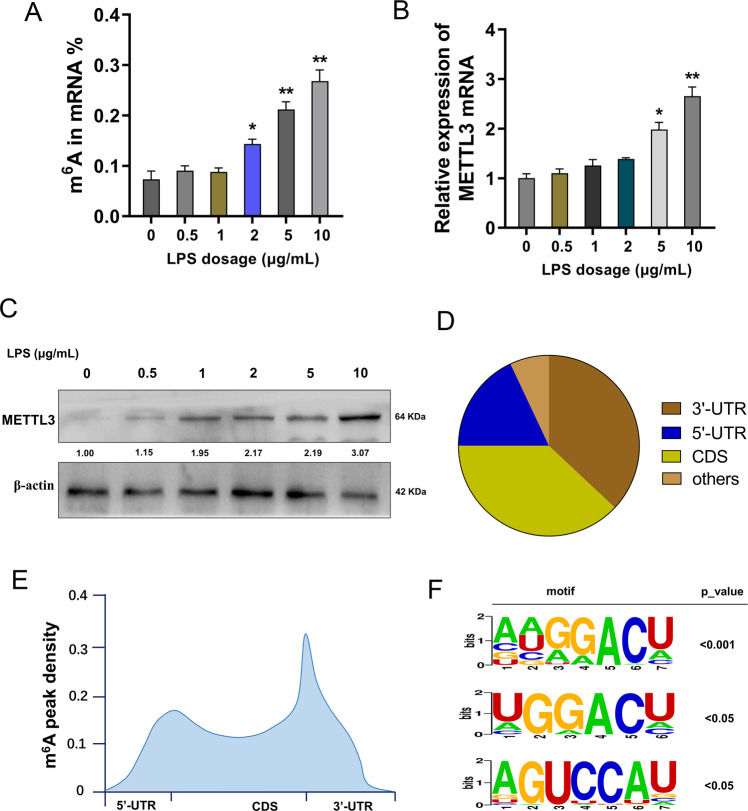
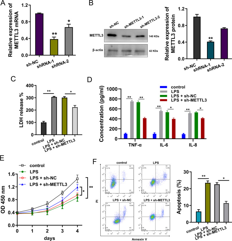
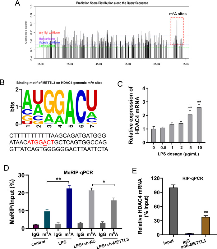
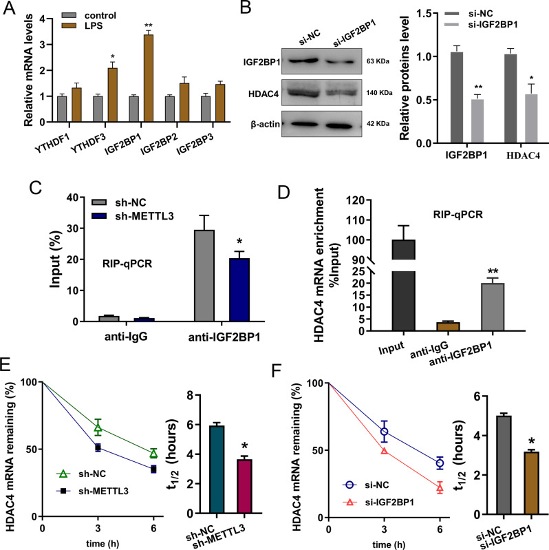
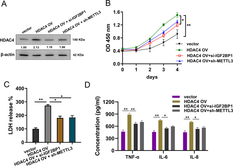
Recent studies have identified that N6-methyladenosine (m6A) extensively participates in the myocardial injury pathophysiological process. However, the role of m6A on sepsis-induced myocardial injury is still unclear. Here, we investigated the functions and mechanism of m6A methyltransferase METTL3 for septic myocardial injury. Results illustrated that the m6A modification level and METTL3 up-regulated in the lipopolysaccharide (LPS)-induced cardiomyocytes (H9C2 cells). Methylated RNA immunoprecipitation sequencing (MeRIP-Seq) revealed the m6A profile of the septic myocardial injury cellular model. Functionally, METTL3 knockdown repressed the inflammatory damage of cardiomyocytes induced by LPS. Mechanistically, we found that HDAC4 had remarkable m6A modification sites on its 3'-UTR genome, acting as the downstream target of METTL3. Besides, m6A reader IGF2BP1 recognized the m6A modification sites on HDAC4 mRNA and enhanced its RNA stability. In conclusion, the findings illustrated a role of METTL3/IGF2BP1/m6A/HDAC4 axis on sepsis-induced myocardial injury, which might provide novel therapeutic strategy for septic myocardial injury.
© 2022. The Author(s).
Conflict of interest statement
The authors declare no competing interests.
Figures
Similar articles
-
N6-methyladenosine writer METTL3 accelerates the sepsis-induced myocardial injury by regulating m6A-dependent ferroptosis.Apoptosis. 2023 Apr;28(3-4):514-524. doi: 10.1007/s10495-022-01808-y. Epub 2023 Jan 16. Apoptosis. 2023. PMID: 36645573
-
N6-Methyladenosine METTL3 Modulates the Proliferation and Apoptosis of Lens Epithelial Cells in Diabetic Cataract.Mol Ther Nucleic Acids. 2020 Jun 5;20:111-116. doi: 10.1016/j.omtn.2020.02.002. Epub 2020 Feb 11. Mol Ther Nucleic Acids. 2020. PMID: 32163892 Free PMC article.
-
METTL3 achieves lipopolysaccharide-induced myocardial injury via m6A-dependent stabilization of Myh3 mRNA.Biochim Biophys Acta Mol Cell Res. 2023 Oct;1870(7):119503. doi: 10.1016/j.bbamcr.2023.119503. Epub 2023 May 27. Biochim Biophys Acta Mol Cell Res. 2023. PMID: 37245538
-
METTL3 plays multiple functions in biological processes.Am J Cancer Res. 2020 Jun 1;10(6):1631-1646. eCollection 2020. Am J Cancer Res. 2020. PMID: 32642280 Free PMC article. Review.
-
Novel insights into the m6A-RNA methyltransferase METTL3 in cancer.Biomark Res. 2021 Apr 20;9(1):27. doi: 10.1186/s40364-021-00278-9. Biomark Res. 2021. PMID: 33879256 Free PMC article. Review.
Cited by
-
Post-translational modifications in sepsis-induced organ dysfunction: mechanisms and implications.Front Immunol. 2024 Aug 21;15:1461051. doi: 10.3389/fimmu.2024.1461051. eCollection 2024. Front Immunol. 2024. PMID: 39234245 Free PMC article. Review.
-
Transcriptome-wide identification of altered RNA m6A profiles in cardiac tissue of rats with LPS-induced myocardial injury.Front Immunol. 2023 May 19;14:1122317. doi: 10.3389/fimmu.2023.1122317. eCollection 2023. Front Immunol. 2023. PMID: 37275860 Free PMC article.
-
An overview of the effects and mechanisms of m6 A methylation on innate immune cells in sepsis.Front Immunol. 2022 Nov 24;13:1041990. doi: 10.3389/fimmu.2022.1041990. eCollection 2022. Front Immunol. 2022. PMID: 36505499 Free PMC article.
-
N6-methyladenosine (m6A) reader IGF2BP1 facilitates clear-cell renal cell carcinoma aerobic glycolysis.PeerJ. 2023 Jan 18;11:e14591. doi: 10.7717/peerj.14591. eCollection 2023. PeerJ. 2023. PMID: 36691477 Free PMC article.
-
RNA m6A methylation regulators in sepsis.Mol Cell Biochem. 2024 Sep;479(9):2165-2180. doi: 10.1007/s11010-023-04841-w. Epub 2023 Sep 2. Mol Cell Biochem. 2024. PMID: 37659034 Review.
References
LinkOut - more resources
Full Text Sources
