. 2022 Jul 15;13(1):4128.
doi: 10.1038/s41467-022-30695-9.

The Medical Segmentation Decathlon

Affiliations

The Medical Segmentation Decathlon

Michela Antonelli et al. Nat Commun. .

Abstract

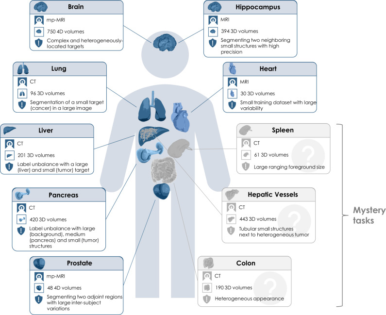
International challenges have become the de facto standard for comparative assessment of image analysis algorithms. Although segmentation is the most widely investigated medical image processing task, the various challenges have been organized to focus only on specific clinical tasks. We organized the Medical Segmentation Decathlon (MSD)-a biomedical image analysis challenge, in which algorithms compete in a multitude of both tasks and modalities to investigate the hypothesis that a method capable of performing well on multiple tasks will generalize well to a previously unseen task and potentially outperform a custom-designed solution. MSD results confirmed this hypothesis, moreover, MSD winner continued generalizing well to a wide range of other clinical problems for the next two years. Three main conclusions can be drawn from this study: (1) state-of-the-art image segmentation algorithms generalize well when retrained on unseen tasks; (2) consistent algorithmic performance across multiple tasks is a strong surrogate of algorithmic generalizability; (3) the training of accurate AI segmentation models is now commoditized to scientists that are not versed in AI model training.


Conflict of interest statement

No funding contributed explicitly to the organization and running of the challenge. The challenge award has been kindly provided by NVIDIA. However, NVIDIA did not influence the design or running of the challenge as they were not part of the organizing committee. R.M.S. received royalties from iCAD, Philips, ScanMed, Translation Holdings, and PingAn. Individually funding sourcing unrelated to the challenge has been listed in the Acknowledgments section. The remaining authors declare no competing interests.

Figures

Fig. 1
Fig. 1. Overview of the ten different tasks of the Medical Segmentation Decathlon (MSD).
The challenge comprised different target regions, modalities and challenging characteristics and was separated into seven known tasks (blue; the development phase: brain, heart, hippocampus, liver, lung, pancreas, prostate) and three mystery tasks (gray; the mystery phase: colon, hepatic vessels, spleen). MRI magnetic resonance imaging, mp-MRI multiparametric-magnetic resonance imaging, CT computed tomography.
Fig. 2
Fig. 2. Base network architectures (left) and loss functions (right) used by the participants of the 2018 Decathlon challenge who provided full algorithmic information (n = 14 teams).
Network architectures: DeepMedic—Efficient multi-scale 3D CNN with fully connected CRF for accurate brain lesion segmentation, QuickNAT—Fully Convolutional Network for Quick and Accurate Segmentation of Neuroanatomy, ResNet—Deep Residual Learning for Image Recognition, U-Net—Convolutional Networks for Biomedical Image Segmentation, V-Net—Fully Convolutional Neural Networks for Volumetric Medical Image Segmentation. DSC Dice Similarity Coefficient.
Fig. 3
Fig. 3. Dot- and box-plots of the Dice Similarity Coefficient (DSC) values of all 19 participating algorithms for the seven tasks (brain, heart, hippocampus, liver, lung, pancreas, prostate) of the development phase, color-coded by the target regions (edema (red), non-enhancing tumor (purple), enhancing tumor (blue), left atrium (green), anterior (olive), posterior (light purple), liver (dark orange), liver tumor (orange), lung tumor (yellow), pancreas (dark yellow), tumor mass (light brown), prostate peripheral zone (PZ) (brown), prostate transition zone (TZ) (pink)).
The box-plots represent descriptive statistics over all test cases. The median value is shown by the black horizontal line within the box, the first and third quartiles as the lower and upper border of the box, respectively, and the 1.5 interquartile range by the vertical black lines. Outliers are shown as black circles. The raw DSC values are provided as gray circles. ROI Region of Interest.
Fig. 4
Fig. 4. Dot- and box-plots of the Dice Similarity Coefficient (DSC) values of all 19 participating algorithms for the three tasks of the mystery phase (colon, hepatic vessel, spleen), color-coded by the target regions (colon cancer primaries (red), hepatic tumor (green), hepatic vessel (yellow), spleen (pink)).
The box-plots represent descriptive statistics over all test cases. The median value is shown by the black horizontal line within the box, the first and third quartiles as the lower and upper border of the box, respectively, and the 1.5 interquartile range by the vertical black lines. Outliers are shown as black circles. The raw DSC values are provided as gray circles. ROI Region of Interest.
Fig. 5
Fig. 5. Dot- and box-plots of ranks for all 19 participating algorithms over all seven tasks and thirteen target regions of the development phase (red) and all three tasks and four target regions of the mystery phase (blue).
The median value is shown by the black vertical line within the box, the first and third quartiles as the lower and upper border of the box, respectively, and the 1.5 interquartile range by the horizontal black lines. Individual ranks are shown as gray circles.

References

    1. Litjens G, et al. Deep learning as a tool for increased accuracy and efficiency of histopathological diagnosis. Sci. Rep. 2016;6:1–11. doi: 10.1038/srep26286. - DOI - PMC - PubMed
    1. Poplin R, et al. Prediction of cardiovascular risk factors from retinal fundus photographs via deep learning. Nat. Biomed. Eng. 2018;2:158–164. doi: 10.1038/s41551-018-0195-0. - DOI - PubMed
    1. Stokes JM, et al. A deep learning approach to antibiotic discovery. Cell. 2020;180:688–702. doi: 10.1016/j.cell.2020.01.021. - DOI - PMC - PubMed
    1. Ayache N, Duncan J. 20th anniversary of the medical image analysis journal (media) Med. Image Anal. 2016;33:1–3. doi: 10.1016/j.media.2016.07.004. - DOI - PubMed
    1. Liang S, et al. Deep-learning-based detection and segmentation of organs at risk in nasopharyngeal carcinoma computed tomographic images for radiotherapy planning. Eur. Radiol. 2019;29:1961–1967. doi: 10.1007/s00330-018-5748-9. - DOI - PubMed

Publication types

MeSH terms