. 2022 Oct;27(10):4035-4049.
doi: 10.1038/s41380-022-01682-9. Epub 2022 Jul 15.

Glucocorticoid-glucocorticoid receptor-HCN1 channels reduce neuronal excitability in dorsal hippocampal CA1 neurons

Affiliations

Glucocorticoid-glucocorticoid receptor-HCN1 channels reduce neuronal excitability in dorsal hippocampal CA1 neurons

Jiwon Kim et al. Mol Psychiatry. 2022 Oct.

Abstract

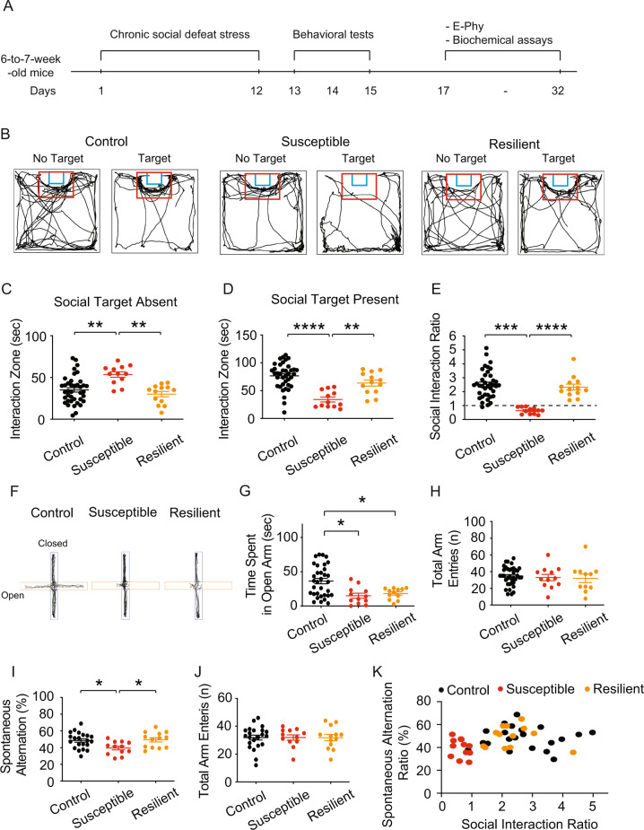
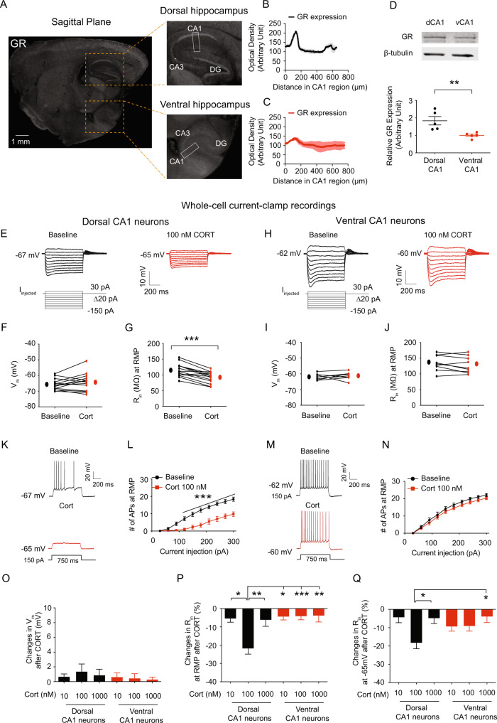
While chronic stress increases hyperpolarization-activated current (Ih) in dorsal hippocampal CA1 neurons, the underlying molecular mechanisms are entirely unknown. Following chronic social defeat stress (CSDS), susceptible mice displayed social avoidance and impaired spatial working memory, which were linked to decreased neuronal excitability, increased perisomatic hyperpolarization-activated cyclic nucleotide-gated (HCN) 1 protein expression, and elevated Ih in dorsal but not ventral CA1 neurons. In control mice, bath application of corticosterone reduced neuronal excitability, increased tetratricopeptide repeat-containing Rab8b-interacting protein (TRIP8b) and HCN1 protein expression, and elevated Ih in dorsal but not ventral CA1 region/neurons. Corticosterone-induced upregulation of functional Ih was mediated by the glucocorticoid receptor (GR), HCN channels, and the protein kinase A (PKA) but not the calcium/calmodulin-dependent protein kinase II (CaMKII) pathway. Three months after the end of CSDS, susceptible mice displayed persistent social avoidance when exposed to a novel aggressor. The sustained behavioral deficit was associated with lower neuronal excitability and higher functional Ih in dorsal CA1 neurons, both of which were unaffected by corticosterone treatment. Our findings show that corticosterone treatment mimics the pathophysiological effects of dorsal CA1 neurons/region found in susceptible mice. The aberrant expression of HCN1 protein along the somatodendritic axis of the dorsal hippocampal CA1 region might be the molecular mechanism driving susceptibility to social avoidance.


Conflict of interest statement

The authors declare no competing interests.

Figures

Fig. 1
Fig. 1. Susceptible mice displayed social avoidance and impaired spatial working memory.
A Timeline of chronic social defeat stress, behavioral tests, electrophysiology, and biochemical assay. B Representative video tracking images of control, susceptible, and resilient mice without and with a social target during the social interaction test. C In the absence of the social target, susceptible group spent more time in the interaction zone than the control and resilient groups. D In the presence of a social target, susceptible group spent less time in the interaction zone than the control and resilient groups. E During the social interaction test, chronic social defeat stress resulted in the formation of susceptible and resilient phenotypes. F Representative video tracking images of age-matched mice during a 6-min elevated plus maze test. G, H The susceptible and resilient groups spent less time in the open arm than the control group without changing the total arm entries. I, J The percentage of spontaneous alternation in susceptible group was much lower than in the control and resilient groups, despite no differences in total arm entries. K The relationship between social avoidance and spatial working memory between groups. Data are expressed as mean ± SEM.
Fig. 2
Fig. 2. The susceptible group reduced Rin and neuronal excitability in dorsal CA1 neurons, but not in ventral CA1 neurons.
AJ We performed whole-cell current-clamp recordings. A representative voltage responses with step current commands ranging from −150  pA to +30 pA (Δ = 20 pA) at RMP. B There was no difference in RMP of dorsal CA1 neurons between groups. C The dorsal CA1 neurons of the susceptible group had lower Rin at RMP than the control and resilient groups. D Representative voltage responses with depolarizing current step (210 pA; 750 ms) at RMP in dorsal CA1 neurons from the control, susceptible, and resilient groups. E Dorsal CA1 neurons from susceptible group had lower action potential firing than the control and resilient groups. F Representative voltage responses with step current commands ranging from −150 pA to +30 pA (Δ = 20 pA) at RMP. G, H There were no differences in RMP and Rin at RMP of ventral CA1 neurons between groups. I Representative voltage responses with depolarizing current step (210 pA; 750 ms) at RMP in the ventral CA1 neurons from the control, susceptible, and resilient groups. J Action potential firing of ventral CA1 neurons was not different between groups. Data are expressed as mean ± SEM.
Fig. 3
Fig. 3. HCN1 protein expression and Ih were significantly increased in dorsal CA1 region/neurons from susceptible group.
A, C Representative dorsal and ventral hippocampal slices immunolabeled with antibody against HCN1. Rectangle boxes depict the region of the slice used for quantification of the optical density. The arrows indicate increased perisomatic HCN1 protein expression. B, D Quantification of HCN1 protein expression from the perisomatic region to the distal dendritic region of CA1 from the dorsal and ventral hippocampi. E Western blot (top) and quantification (bottom) of HCN1 protein in dorsal CA1 region from the control, susceptible, and resilient groups. F, G We performed cell-attached voltage-clamp recordings. F Representative maximal h current traces in response to a 500-ms hyperpolarizing voltage step (−140 mV). G Ih was significantly increased in dorsal CA1 neurons from susceptible group compared with those from the control and resilient groups. HL We performed whole-cell voltage-clamp recordings. H Representative current responses with step voltage commands ranging from −140 mV to − 60 mV (Δ = 10 mV) at a holding potential of −60 mV. The approximate position for determining the peak tail current is shown by black vertical dashed lines. I Susceptible group showed increased Ih in the dorsal CA1 neurons compared with those from the control and resilient groups. J The voltage dependence of activation for h channel was determined from tail currents (Ih / Ih max). The activation curve was fitted with a Boltzmann function with the following values: control V1/2 = −101.8 mV, k = −14.13 mV, susceptible V1/2 = −90.02 mV, k = −16.09 mV, resilient V1/2 = −102.1 mV, k = −19.9 mV. K The half-activation voltage of h channel (V1/2) for susceptible group was significantly shifted to the right by around +10 mV, whereas L the slope factor was not different between groups. Data are expressed as mean ± SEM.
Fig. 4
Fig. 4. Dorsal CA1 neurons responded to corticosterone more strongly than ventral CA1 neurons.
A Representative sagittal section of brain showing GR immunoreactivity in dorsal (top) and ventral (bottom) hippocampi. Rectangle boxes depict the region of the slice used for quantification of optical density. B, C Quantification of GR protein expression from the perisomatic region to the distal dendritic region of CA1 from the dorsal and ventral hippocampi. D Western blot (top) and quantification (bottom) of GR protein in CA1 region from the dorsal and ventral hippocampi. EQ We performed whole-cell current-clamp recordings. E, H Representative voltage responses with step current commands ranging from −150 pA to +30 pA (Δ = 20 pA) at RMP before and after bath application of corticosterone. Corticosterone lowered Rin (G) but had no impact on RMP (F) in dorsal CA1 neurons. There are no changes in RMP (I) and Rin (J) in ventral CA1 neurons following corticosterone treatment. Representative voltage responses with depolarizing current step (150 pA; 750 ms) at RMP in dorsal (K) and ventral (M) CA1 neurons. L Dorsal CA1 neurons had decreased action potential firing at RMP following corticosterone treatment. N Action potential firing at RMP was not altered in ventral CA1 neurons following corticosterone treatment. O Corticosterone doses of 10 nM, 100 nM, and 1 μM had no effect on RMP of CA1 neurons in dorsal and ventral hippocampi. 100 nM corticosterone significantly reduced Rin at RMP (P) and at −65 mV (Q) in dorsal CA1 neurons compared to 10 nM and 1 μM corticosterone. When compared to 10 nM and 1 μM corticosterone, 100 nM corticosterone had no impact on Rin at RMP (P) and at −65 mV (Q) in ventral CA1 neurons. Data are expressed as mean ± SEM.
Fig. 5
Fig. 5. The GR, HCN channels, and the PKA pathway were all involved in corticosterone-induced upregulation of functional Ih.
A Illustration depicting the acute corticosterone treatment in the dorsal hippocampus. B Representative dorsal hippocampal slices immunolabeled with antibody against TRIP8b. Rectangle boxes depict the region of the slice used for quantification of the optical density. The arrows indicate increased perisomatic TRIP8b protein expression. C Quantification of TRIP8b protein expression from the perisomatic region to the distal dendritic region of CA1 from the dorsal hippocampus. D Representative dorsal hippocampal slices immunolabeled with antibody against HCN1. Rectangle boxes depict the region of the slice used for quantification of the optical density. The arrows indicate increased perisomatic HCN1 protein expression. E Quantification of HCN1 protein expression from the perisomatic region to the distal dendritic region of CA1 from the dorsal hippocampus. F, G We performed cell-attached voltage-clamp recordings. F Representative maximal h current traces in response to a 500-ms hyperpolarizing voltage step (−140 mV). G Ih was significantly elevated in the dorsal CA1 neurons from corticosterone treatment compared with those from the vehicle-treated group. Dexamethasone reduced Rin at RMP (H) and at −65 mV (J) in dorsal CA1 neurons. Corticosterone-induced decrease in Rin at RMP (H) and at −65 mV (J) was blocked by RU 486, KT5720, and ZD7288. Changes in Rin at RMP (I) and at −65 mV (K) were blocked by RU 486, KT5720, and ZD7288. Data are expressed as mean ± SEM.
Fig. 6
Fig. 6. Susceptible mice re-exposed to novel aggressor mice displayed a persistent social avoidance, which was associated with elevated Ih and was insensitive to corticosterone effects.
A Timeline of CSDS, behavioral tests, electrophysiology, and biochemical assay. B CSDS produced the susceptible and resilient phenotype during the social interaction test. After 1 month (C) or 3 months (D) of no CSDS, susceptible mice showed persistent social avoidance during the social interaction test. E, F We performed whole-cell current-clamp recordings. E Representative voltage responses with depolarizing current step (210 pA; 750 ms) at RMP in dorsal CA1 neurons. F Dorsal CA1 neurons of susceptible group had lower action potential firing than control group, whereas the resilient group had higher action potential firing. G, H We performed cell-attached voltage-clamp recordings. G Representative maximal h current traces in response to a 500-ms hyperpolarizing voltage step (−140 mV). H Ih was significantly elevated in the dorsal CA1 neurons from susceptible group compared with those from the control and resilient mice. Representative dorsal hippocampal slices immunolabeled with antibody against HCN1 (I) and TRIP8b (K). Rectangle boxes depict the region of the slice used for quantification of the optical density. The arrows indicate increased perisomatic HCN1 and TRIP8b protein expression. Quantification of HCN1 (J) and TRIP8b (L) protein expression from the perisomatic region to the distal dendritic region of CA1 from the dorsal hippocampus. M, N We performed whole-cell current-clamp recordings. M Corticosterone reduced Rin at RMP of the dorsal CA1 neurons in the control and resilient groups, but had no effect on Rin in susceptible group. N Changes in Rin at RMP were much lower in susceptible group compared to the control and resilient groups. Data are expressed as mean ± SEM.

Similar articles

Cited by

References

    1. American_Psychiatric_Association. Diagnostic and Statistical Manual of Mental Disorders 5th Edn. Washington, DC: American Psychiatric Association; 2013.
    1. Kessler RC. Posttraumatic stress disorder: the burden to the individual and to society. J Clin Psychiatry. 2000;61:4–12. - PubMed
    1. Buckley TC, Mozley SL, Bedard MA, Dewulf AC, Greif J. Preventive health behaviors, health-risk behaviors, physical morbidity, and health-related role functioning impairment in veterans with post-traumatic stress disorder. Mil Med. 2004;169:536–40. - PubMed
    1. Haviland MG, Banta JE, Sonne JL, Przekop P. Posttraumatic Stress Disorder-Related Hospitalizations in the United States (2002-11): Rates, Co-Occurring Illnesses, Suicidal Ideation/Self-Harm, and Hospital Charges. J Nerv Ment Dis. 2016;204:78–86. - PubMed
    1. Rytwinski NK, Scur MD, Feeny NC, Youngstrom EA. The co-occurrence of major depressive disorder among individuals with posttraumatic stress disorder: a meta-analysis. J Trauma Stress. 2013;26:299–309. - PubMed

Publication types

MeSH terms

Substances