

Editorial
doi: 10.1007/s00259-022-05913-7.
2-[18F]-FDG PET for imaging brain involvement in patients with long COVID: perspective of the EANM Neuroimaging Committee
Affiliations
- PMID: 35840817
- PMCID: PMC9286958
- DOI: 10.1007/s00259-022-05913-7
Item in Clipboard
Editorial
2-[18F]-FDG PET for imaging brain involvement in patients with long COVID: perspective of the EANM Neuroimaging Committee
Eur J Nucl Med Mol Imaging.
2022 Sep.
No abstract available
Conflict of interest statement
AV has received speaker honoraria from GE Healthcare and Philips.
HB has received reader honoraria from LMI and speaker honoraria from AAA/Novartis.
FF has received speaker honoraria and receives research funding from Siemens Healthineers.
SM has received speaker honoraria from GE Healthcare, Roche, and LMI and is an advisor of LMI.
EG has received consultant and speaker honoraria from GE Healthcare and CIS Bio International; and consultant honoraria from Advanced Accelerator Applications
The other authors declare that they have no conflict of interest
Figures
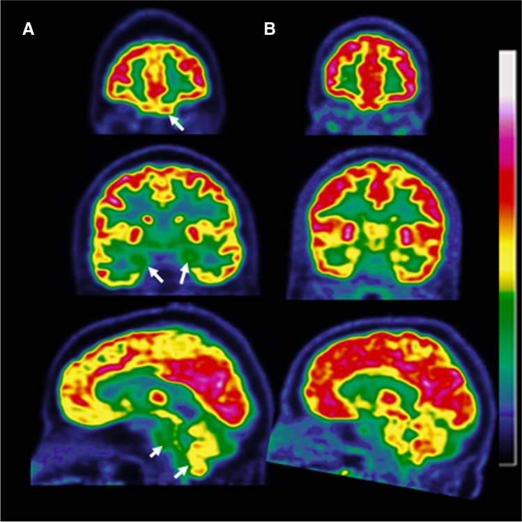
Hypometabolic pattern in a patient with long COVID-19 presenting normal MRI (A) and normal brain FDG PET scans of an age-matched control subject (B). The hypometabolic long COVID pattern involves the fronto-orbital and olfactory regions and other limbic/paralimbic regions as visualized in coronal slices, and the brainstem and the cerebellum as visualized in sagittal slices (white arrows in A)
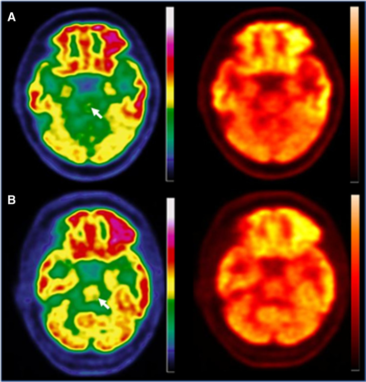
Axial slices of a brain FDG PET scan of a patient with a hypometabolic long COVID pattern (A) and in a patient with a normal brain scan (B). A discontinuous colour scale (left column) is better suited than a continuous scale (right column) to indicate hypometabolism in the pons (white arrow)
References
Publication types
MeSH terms
Substances
LinkOut - more resources
Full Text Sources
Medical
