

Morphologic CT and MRI features of primary parotid squamous cell carcinoma and its predictive factors for differential diagnosis with mucoepidermoid carcinoma
- PMID: 35840821
- PMCID: PMC9287497
- DOI: 10.1186/s13244-022-01256-x
Morphologic CT and MRI features of primary parotid squamous cell carcinoma and its predictive factors for differential diagnosis with mucoepidermoid carcinoma
Abstract
Background: Primary parotid squamous cell carcinoma (SCC) is a rare entity with a poor prognosis. Pathologically, the diagnosis of it requires the exclusion of parotid mucoepidermoid carcinoma (MEC). Currently, the imaging features of primary parotid SCC and the predictive indicators for differential diagnosis of the two entities have not been well reported. Our purpose was to identify the imaging characteristics of primary parotid SCC and to determine the predictive factors for its' differential diagnosis.
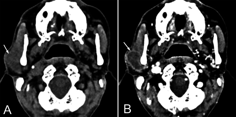
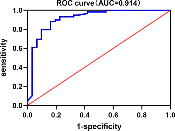
Results: Thirty-one participants with primary parotid SCC and 59 with primary parotid MEC were enrolled. Clinical, CT and MRI features were reviewed and compared by univariate analysis. Then, multinomial logistic regression was used to determine the predictors to distinguish parotid SCC from MEC. Most primary parotid SCCs exhibited irregular shape, ill-defined margin, incomplete or no capsule, heterogeneous and marked or moderate enhancement, necrosis, local tumor invasiveness (LTI). Age, maximal dimension, shape, degree of enhancement, gradual enhancement, necrosis, and LTI were different between the primary parotid SCCs and MECs in univariate analysis (p < 0.05). While in multinomial logistic regression analysis, only age and necrosis were the independent predictors for distinguishing parotid SCC from MEC, and this model exhibited an area under curve of 0.914 in ROC curve analysis.
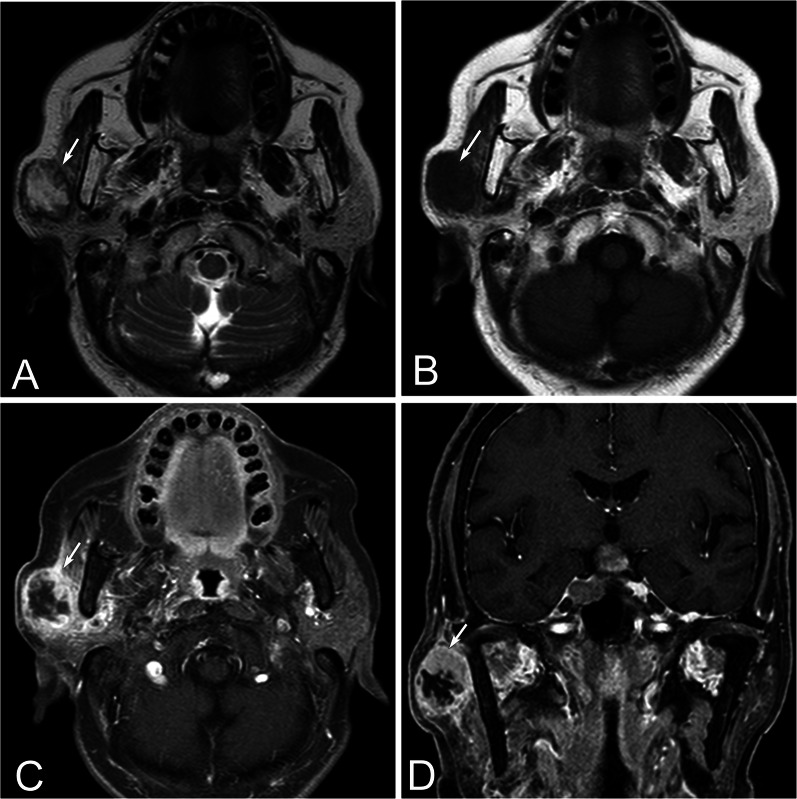
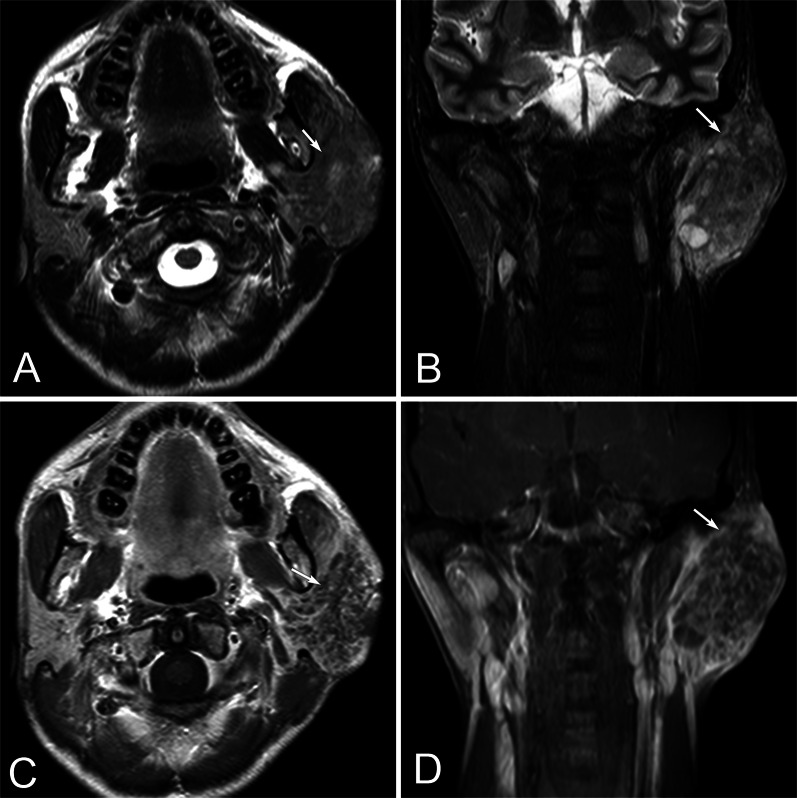
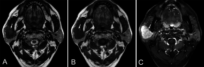
Conclusions: Primary parotid SCC has some distinct imaging features including the large tumor size, irregular shape, ill-defined margin, and particularly the marked central necrosis. Patients with age ≥ 51.5 years and necrosis on the image of the primary tumor in the parotid gland could be more likely to be SCCs than MECs.
Keywords: Diagnosis; Imaging; Parotid gland; Predictive; Primary; Squamous cell carcinomas.
© 2022. The Author(s).
Conflict of interest statement
All the authors declare that they have no competing interests.
Figures
Similar articles
-
Predictive CT features for the diagnosis of primary pulmonary mucoepidermoid carcinoma: comparison with squamous cell carcinomas and adenocarcinomas.Cancer Imaging. 2021 Jan 6;21(1):2. doi: 10.1186/s40644-020-00375-2. Cancer Imaging. 2021. PMID: 33407915 Free PMC article.
-
[Value of manifestations of magnetic resonance imaging (MRI) in diagnosis of parotid tumors and their pathological bases].Ai Zheng. 2003 May;22(5):514-9. Ai Zheng. 2003. PMID: 12753715 Chinese.
-
Squamous cell carcinoma originating in the parotid gland: MRI features with histopathological correlation.Clin Radiol. 2014 Jan;69(1):41-4. doi: 10.1016/j.crad.2013.08.002. Epub 2013 Sep 30. Clin Radiol. 2014. PMID: 24090910
-
Understanding primary parotid squamous cell carcinoma - A systematic review.Surgeon. 2020 Feb;18(1):44-48. doi: 10.1016/j.surge.2019.03.006. Epub 2019 Apr 28. Surgeon. 2020. PMID: 31040083
-
Warthin-Like Mucoepidermoid Carcinoma: A Morphological Spectrum - A Report of 3 Cases with Histological and Cytological Findings and Review of the Literature.Acta Cytol. 2022;66(3):244-252. doi: 10.1159/000521134. Epub 2022 Feb 4. Acta Cytol. 2022. PMID: 35124667 Review.
Cited by
-
A deep learning-based radiomics model for predicting lymph node status from lung adenocarcinoma.BMC Med Imaging. 2024 May 24;24(1):121. doi: 10.1186/s12880-024-01300-w. BMC Med Imaging. 2024. PMID: 38789936 Free PMC article.
-
MRI and CT imaging characteristics in parotid tumors with false-negative fine-needle aspirations.Head Face Med. 2024 Oct 26;20(1):64. doi: 10.1186/s13005-024-00467-5. Head Face Med. 2024. PMID: 39462423 Free PMC article.
-
A Cautionary Tale: A Case Report Describing a Benign Parotid Oncocytoma Diagnosed as Metastatic Squamous Cell Carcinoma on Fine Needle Aspirate.Cureus. 2023 Dec 20;15(12):e50853. doi: 10.7759/cureus.50853. eCollection 2023 Dec. Cureus. 2023. PMID: 38249277 Free PMC article.
References
LinkOut - more resources
Full Text Sources
Research Materials
