

Use data augmentation for a deep learning classification model with chest X-ray clinical imaging featuring coal workers' pneumoconiosis
- PMID: 35840945
- PMCID: PMC9284687
- DOI: 10.1186/s12890-022-02068-x
Use data augmentation for a deep learning classification model with chest X-ray clinical imaging featuring coal workers' pneumoconiosis
Abstract
Purpose: This paper aims to develop a successful deep learning model with data augmentation technique to discover the clinical uniqueness of chest X-ray imaging features of coal workers' pneumoconiosis (CWP).
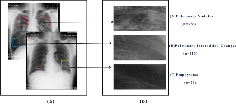
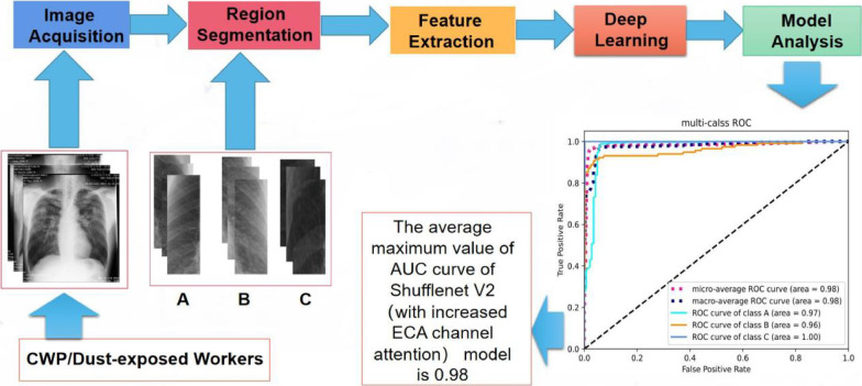
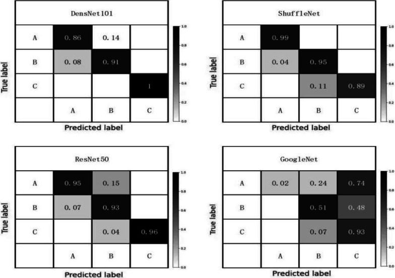
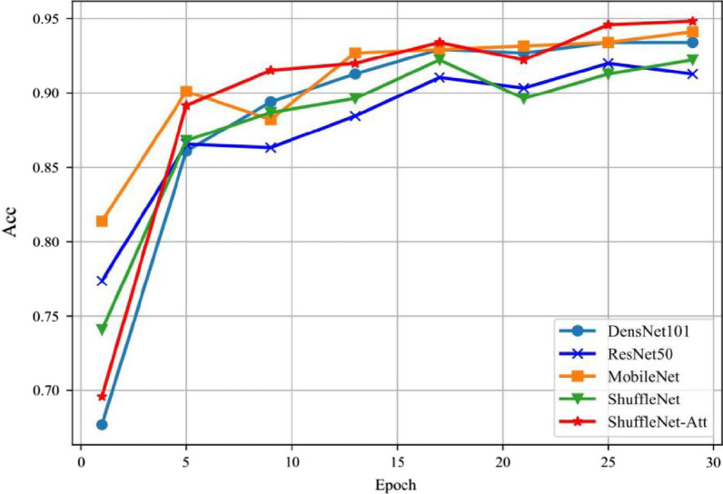
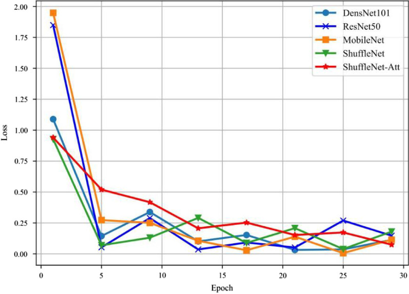
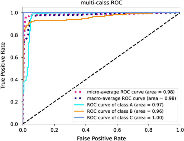
Patients and methods: We enrolled 149 CWP patients and 68 dust-exposure workers for a prospective cohort observational study between August 2021 and December 2021 at First Hospital of Shanxi Medical University. Two hundred seventeen chest X-ray images were collected for this study, obtaining reliable diagnostic results through the radiologists' team, and confirming clinical imaging features. We segmented regions of interest with diagnosis reports, then classified them into three categories. To identify these clinical features, we developed a deep learning model (ShuffleNet V2-ECA Net) with data augmentation through performances of different deep learning models by assessment with Receiver Operation Characteristics (ROC) curve and area under the curve (AUC), accuracy (ACC), and Loss curves.
Results: We selected the ShuffleNet V2-ECA Net as the optimal model. The average AUC of this model was 0.98, and all classifications of clinical imaging features had an AUC above 0.95.
Conclusion: We performed a study on a small dataset to classify the chest X-ray clinical imaging features of pneumoconiosis using a deep learning technique. A deep learning model of ShuffleNet V2 and ECA-Net was successfully constructed using data augmentation, which achieved an average accuracy of 98%. This method uncovered the uniqueness of the chest X-ray imaging features of CWP, thus supplying additional reference material for clinical application.
Keywords: Chest X-ray; Coal workers' pneumoconiosis classification; Data augmentation; Deep learning; ECA-Net; ShuffleNet.
© 2022. The Author(s).
Conflict of interest statement
The authors declare that they have no competing interests.
Figures
References
Publication types
MeSH terms
Substances
Grants and funding
- No.2020-PT320-005/The National Health Commission Key Laboratory of Pneumoconiosis Shanxi China Project
- No.2020-PT320-005/The National Health Commission Key Laboratory of Pneumoconiosis Shanxi China Project
- No.2020-PT320-005/The National Health Commission Key Laboratory of Pneumoconiosis Shanxi China Project
- No.2020-PT320-005/The National Health Commission Key Laboratory of Pneumoconiosis Shanxi China Project
LinkOut - more resources
Full Text Sources
