. 2022 Jul 15;19(1):48.
doi: 10.1186/s12989-022-00490-x.

Titanium dioxide and carbon black nanoparticles disrupt neuronal homeostasis via excessive activation of cellular prion protein signaling

Affiliations

Titanium dioxide and carbon black nanoparticles disrupt neuronal homeostasis via excessive activation of cellular prion protein signaling

Luiz W Ribeiro et al. Part Fibre Toxicol. .

Abstract

Background: Epidemiological emerging evidence shows that human exposure to some nanosized materials present in the environment would contribute to the onset and/or progression of Alzheimer's disease (AD). The cellular and molecular mechanisms whereby nanoparticles would exert some adverse effects towards neurons and take part in AD pathology are nevertheless unknown.

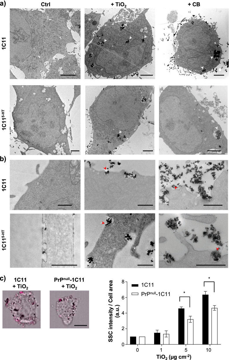
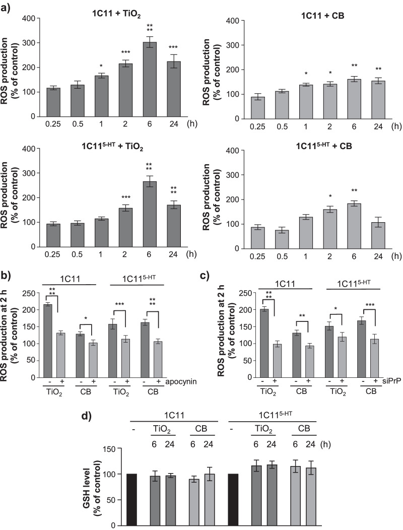
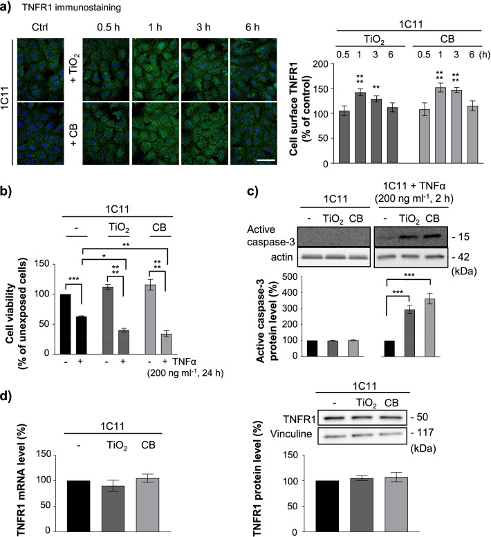
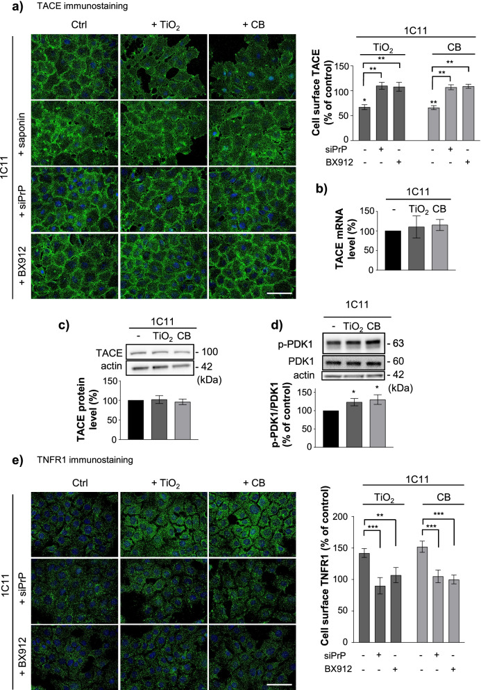
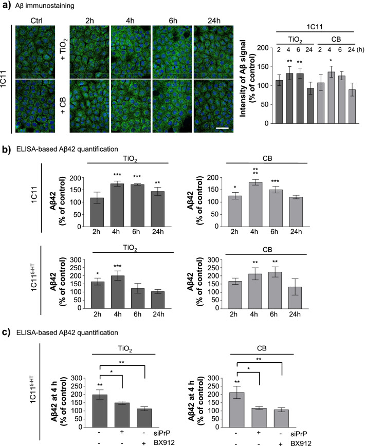
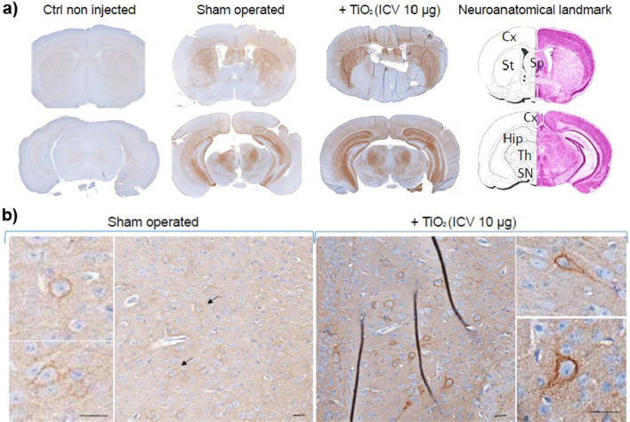
Results: Here, we provide the prime evidence that titanium dioxide (TiO2) and carbon black (CB) nanoparticles (NPs) bind the cellular form of the prion protein (PrPC), a plasma membrane protein well known for its implication in prion diseases and prion-like diseases, such as AD. The interaction between TiO2- or CB-NPs and PrPC at the surface of neuronal cells grown in culture corrupts PrPC signaling function. This triggers PrPC-dependent activation of NADPH oxidase and subsequent production of reactive oxygen species (ROS) that alters redox equilibrium. Through PrPC interaction, NPs also promote the activation of 3-phosphoinositide-dependent kinase 1 (PDK1), which in turn provokes the internalization of the neuroprotective TACE α-secretase. This diverts TACE cleavage activity away from (i) TNFα receptors (TNFR), whose accumulation at the plasma membrane augments the vulnerability of NP-exposed neuronal cells to TNFα -associated inflammation, and (ii) the amyloid precursor protein APP, leading to overproduction of neurotoxic amyloid Aβ40/42 peptides. The silencing of PrPC or the pharmacological inhibition of PDK1 protects neuronal cells from TiO2- and CB-NPs effects regarding ROS production, TNFα hypersensitivity, and Aβ rise. Finally, we show that dysregulation of the PrPC-PDK1-TACE pathway likely occurs in the brain of mice injected with TiO2-NPs by the intra-cerebro-ventricular route as we monitor a rise of TNFR at the cell surface of several groups of neurons located in distinct brain areas.

Conclusion: Our in vitro and in vivo study thus posits for the first time normal cellular prion protein PrPC as being a neuronal receptor of TiO2- and CB-NPs and identifies PrPC-coupled signaling pathways by which those nanoparticles alter redox equilibrium, augment the intrinsic sensitivity of neurons to neuroinflammation, and provoke a rise of Aβ peptides. By identifying signaling cascades dysregulated by TiO2- and CB-NPs in neurons, our data shed light on how human exposure to some NPs might be related to AD.

Keywords: Alzheimer’s disease; Aβ peptides; Nanoneurotoxicity; Nanoparticles; Neuroinflammation; PrPC receptor; Signaling; TNFα receptors.


Conflict of interest statement

The authors declare that they have no competing interests.

Figures

Fig. 1
Fig. 1
TiO2 and CB nanoparticles interact with PrPC. a Schematic of the experimental procedure to assess the interaction between full-length recombinant mouse PrPC (recPrPC, 2 µM) and TiO2- or CB-NPs (0–80 µg ml−1) exploiting the intrinsic fluorescence of PrPC or by western blotting (WB). b Fluorescence titration curves showing the binding of TiO2- or CB-NPs to full-length mouse recPrPC. The quantity of recPrPC bound with nanoparticles was deduced by subtracting the fluorescence level of titrated free PrPC to the fluorescence level of total PrPC measured in the absence of nanoparticles. Fitting hyperbolic curves were calculated with the help of the Kaleidagraph Software (Abelbeck Software). c Representative Western-blot and quantification histogram showing decrease of free recPrPC amount in the supernatant of the centrifuged reaction medium between recPrPC and TiO2- or CB-NPs. The experiments were performed three times in triplicates. Values are means ± SEM. *denotes p < 0.05, ***p < 0.001, ****p < 0.0001 versus recPrPC incubated without nanoparticles
Fig. 2
Fig. 2
PrPC facilitates nanoparticle interaction with plasma membrane of 1C11 precursors and 1C115−HT neuronal cells. a, b Transmission Electron Microscopy experiments showing large aggregates of TiO2- and CB-NPs (white arrows) within 1C11 precursor cells and 1C115−HT neuronal cells a and small aggregates of TiO2- and CB-NPs (red arrows) at the plasma membrane of 1C11 and 1C115−HT cells b after 24 h exposure to 10 µg cm−2 nanoparticles. Scale bar = 2 µm in a. Scale bar = 0.5 µm in b. c Light scattering-based FACS analysis of interacting TiO2-NPs with the surface of 1C11 and PrPnull-1C11 cells exposed up to 10 µg cm−2 nanoparticles. (Left) Representative bright-field images of 1C11 and PrPnull-1C11 cells exposed to 5 µg cm−2 TiO2-NPs merged with the light scattering signal of TiO2-NPs (pink signal) adsorbed to the plasma membrane after internal masking of cells (see Methods). (Right) Quantification histogram of TiO2-NPs present at the surface of 1C11 and PrPnull-1C11 cells. Scale bar = 5 µm. The experiments were performed three times in triplicates. Values are means ± SEM. *denotes p < 0.05 versus 1C11 cells exposed to nanoparticles
Fig. 3
Fig. 3
TiO2- and CB-NPs specifically interact with full-length PrPC in the 1C11 neuronal cell line. a Schematic of the experimental procedure to assess the interaction of TiO2- and CB-NPs with PrPC expressed at the plasma membrane of 1C11 precursor cells and their serotonergic 1C115−HT neuronal progenies by western blotting. b Quantification histogram deduced from Western-blot experiments showing the amounts of full-length (FL) PrPC (left) and C1 fragment (right) in the supernatant (S fraction) obtained after lysis of 1C11 cells exposed to increasing concentrations of TiO2- or CB-NPs (0 to 10 µg cm−2) for 15 min and centrifugation of the lysates. c, d Representative Western-blots and quantification histograms showing time-variation in the level of FL PrPC and C1 fragment in the S and P fractions derived from 1C11 c and 1C115−HT d cells exposed to TiO2- or CB-NPs (1 µg cm−2). α-tubulin was used for normalization of PrPC level in the S fraction. The experiments were performed three times in triplicates. Values are means ± SEM. *denotes p < 0.05, **p < 0.01, ***p < 0.001, ****p < 0.0001 versus unexposed cells
Fig. 4
Fig. 4
TiO2 and CB nanoparticle interaction with PrPC promotes NADPH oxidase-dependent ROS production. a Kinetics of ROS production induced on exposure to TiO2- and CB-NPs (1 µg cm−2) in 1C11 and 1C115−HT neuronal cells. b Involvement of NADPH oxidase in NP-induced ROS production (1 µg cm−2 for 2 h) using the NADPH oxidase inhibitor apocynin (500 µM). c Quantification histogram showing that siRNA-based PrPC silencing (siPrP) abrogates ROS production induced by TiO2- or CB-NPs (1 µg cm−2 for 2 h) in 1C11 and 1C115−HT cells. d GSH level keeps constant in 1C11 and 1C115−HT neuronal cells exposed to TiO2- or CB-NPs (1 µg cm−2) up to 24 h. The experiments were performed three times in triplicates. Values are means ± SEM. *denotes p < 0.05, **p < 0.01, ***p < 0.001, ****p < 0.0001 versus unexposed cells
Fig. 5
Fig. 5
TiO2 and CB nanoparticle interaction with PrPC renders 1C11 and 1C115−HT neuronal cells highly sensitive to TNFα insult by promoting TNFR1 accumulation at the plasma membrane. a Kinetics of TNFR1 rise at the plasma membrane of 1C11 cells exposed to TiO2- or CB-NPs (1 µg cm−2) and related quantification histogram of immunostained TNFR1. Scale bar = 10 µm. b Viability and c caspase-3 activation in 1C11 cells exposed to TiO2- or CB-NPs (1 µg cm−2) in combination or not with TNFα (200 ng ml−1). d TNFR1 expression level as assessed by RT-qPCR and Western-blotting in 1C11 cells exposed to TiO2- or CB-NPs (1 µg cm−2) for 1 h. Vinculine was used for normalization. The experiments were performed three times in triplicates. Values are means ± SEM. *denotes p < 0.05, **p < 0.01, ***p < 0.001, ****p < 0.0001 versus unexposed cells
Fig. 6
Fig. 6
TiO2 and CB nanoparticle interaction with PrPC promotes TACE internalization in a PDK1-dependent manner at the root of TNFR1 overexposure at the cell surface. a TACE immunostaining at the plasma membrane of 1C11 cells exposed to TiO2- or CB-NPs (1 µg cm−2) for 1 h in the presence or not of a siRNA toward PrPC (siPrP) or the PDK1 inhibitor, BX912 (1 µM) and related quantification histogram. Cell permeabilization with saponin (0.05%) shows TACE internalization in 1C11 cells exposed to nanoparticles. Scale bar = 10 µm. b, c TACE expression level as assessed by RT-qPCR b and Western-blotting c in 1C11 cells exposed to TiO2- or CB-NPs (1 µg cm−2) for 1 h. d PDK1 phosphorylation status at Ser241 (p-PDK1) was assessed by Western-blotting in 1C11 cells exposed to TiO2- or CB-NPs (1 µg cm−2) for 1 h. e TNFR1 immunostaining at the plasma membrane of 1C11 cells exposed for 1 h to TiO2- or CB-NPs (1 µg cm−2) in the presence or not of a siRNA toward PrPC (siPrP) or the PDK1 inhibitor, BX912 (1 µM) and related quantification histogram. Scale bar = 10 µm. The experiments were performed three times in triplicates. Values are means ± SEM. *denotes p < 0.05, **p < 0.01, ***p < 0.001 versus unexposed cells
Fig. 7
Fig. 7
Cell exposure to TiO2 and CB nanoparticles promotes rise of Aβ42 through corruption of the PrPC-PDK1 pathway. a Kinetics of intracellular Aβ accumulation in 1C11 cells exposed to TiO2- or CB-NPs (1 µg cm−2) and related quantification histogram of immunostained Aβ. Scale bar = 10 µm. b ELISA-based quantification of Aβ42 peptides in 1C11 and 1C115−HT neuronal cells exposed to TiO2- or CB-NPs (1 µg cm−2) up to 24 h. c ELISA-based quantification of Aβ42 peptides in 1C115−HT neuronal cells exposed to TiO2- or CB-NPs (1 µg cm−2) for 4 h in the presence or not of a siRNA toward PrPC (siPrP) or the PDK1 inhibitor, BX912 (1 µM). The experiments were performed three times in triplicates. Values are means ± SEM. *denotes p < 0.05, **p < 0.01, ***p < 0.001, ****p < 0.0001 versus unexposed cells
Fig. 8
Fig. 8
TiO2 nanoparticles trigger TNFR1 accumulation in vivo. a TNFR1 immunostaining on two representative brain sections from non-injected, sham-operated, and mice exposed to 10 µg of TiO2-NPs delivered by an unilateral intra-cerebro-ventricular (ICV) injection 8 weeks before. TNFR1 was detected in the cortex (Cx), septum (Sp) striatum (St), hippocampus (Hip), thalamic (Th) nuclei, and substantia nigra (SN). b Higher magnification of the subicular cortex shows TNFR1 rise at the plasma membrane of neurons that is stronger in TiO2-NPs-injected mice than in sham-operated mice (arrows). Scale bar = 25 µm

Similar articles

Cited by

References

    1. Bencsik A, Lestaevel P, Guseva CI. Nano- and neurotoxicology: an emerging discipline. Prog Neurobiol. 2018;160:45–63. doi: 10.1016/j.pneurobio.2017.10.003. - DOI - PubMed
    1. Kaphle A, Navya PN, Umapathi A, Daima HK. Nanomaterials for agriculture, food and environment: applications, toxicity and regulation. Environ Chem Lett. 2018;16:43–58. doi: 10.1007/s10311-017-0662-y. - DOI
    1. Wu T, Tang M. Review of the effects of manufactured nanoparticles on mammalian target organs. J Appl Toxicol. 2018;38:25–40. doi: 10.1002/jat.3499. - DOI - PubMed
    1. Deuschl G, Beghi E, Fazekas F, Varga T, Christoforidi KA, Sipido E, et al. The burden of neurological diseases in Europe: an analysis for the global burden of disease study 2017. Lancet Public Health Elsevier. 2020;5:e551–e567. doi: 10.1016/S2468-2667(20)30190-0. - DOI - PubMed
    1. De Strooper B, Vassar R, Golde T. The secretases: enzymes with therapeutic potential in Alzheimer disease. Nat Rev Neurol. 2010;6:99–107. doi: 10.1038/nrneurol.2009.218. - DOI - PMC - PubMed

Publication types