. 2022 Jul 15;13(1):315.
doi: 10.1186/s13287-022-02995-w.

Exosomal miR-132-3p from mesenchymal stromal cells improves synaptic dysfunction and cognitive decline in vascular dementia

Affiliations

Exosomal miR-132-3p from mesenchymal stromal cells improves synaptic dysfunction and cognitive decline in vascular dementia

Xiaotang Ma et al. Stem Cell Res Ther. .

Abstract

Background/aims: Vascular dementia (VD) results in cognition and memory deficit. Exosomes and their carried microRNAs (miRs) contribute to the neuroprotective effects of mesenchymal stromal cells, and miR-132-3p plays a key role in neuron plasticity. Here, we investigated the role and underlying mechanism of MSC EX and their miR-132-3p cargo in rescuing cognition and memory deficit in VD mice.

Methods: Bilateral carotid artery occlusion was used to generate a VD mouse model. MiR-132-3p and MSC EX levels in the hippocampus and cortex were measured. At 24-h post-VD induction, mice were administered with MSC EX infected with control lentivirus (EXCon), pre-miR-132-3p-expressing lentivirus (EXmiR-132-3p), or miR-132-3p antago lentivirus (EXantagomiR-132-3p) intravenously. Behavioral and cognitive tests were performed, and the mice were killed in 21 days after VD. The effects of MSC EX on neuron number, synaptic plasticity, dendritic spine density, and Aβ and p-Tau levels in the hippocampus and cortex were determined. The effects of MSC EX on oxygen-glucose deprivation (OGD)-injured neurons with respect to apoptosis, and neurite elongation and branching were determined. Finally, the expression levels of Ras, phosphorylation of Akt, GSK-3β, and Tau were also measured.

Results: Compared with normal mice, VD mice exhibited significantly decreased miR-132-3p and MSC EX levels in the cortex and hippocampus. Compared with EXCon treatment, the infusion of EXmiR-132-3p was more effective at improving cognitive function and increasing miR-132-3p level, neuron number, synaptic plasticity, and dendritic spine density, while decreasing Aβ and p-Tau levels in the cortex and hippocampus of VD mice. Conversely, EXantagomiR-132-3p treatment significantly decreased miR-132-3p expression in cortex and hippocampus, as well as attenuated EXmiR-132-3p treatment-induced functional improvement. In vitro, EXmiR-132-3p treatment inhibited RASA1 protein expression, but increased Ras and the phosphorylation of Akt and GSK-3β, and decreased p-Tau levels in primary neurons by delivering miR-132-3p, which resulted in reduced apoptosis, and increased neurite elongation and branching in OGD-injured neurons.

Conclusions: Our studies suggest that miR-132-3p cluster-enriched MSC EX promotes the recovery of cognitive function by improving neuronal and synaptic dysfunction through activation of the Ras/Akt/GSK-3β pathway induced by downregulation of RASA1.

Keywords: Exosomes; Mesenchymal stromal cells; Synaptic plasticity; Vascular dementia; miR-132-3p.


Conflict of interest statement

The authors declare that they have no competing interests.

Figures

Fig. 1
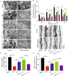
Fig. 1
EXmiR-132-3p infusion restores cortical and hippocampal MSC EX and miR-132-3p level in VD mice. AC MSC EX from sham and VD mouse brain tissue was detected by NTA, TEM, and western blotting. D The effects of EXCon/EXmiR-132-3p/EXantagomiR-132-3p infusion on cortical and hippocampal MSC EX level in Sham and VD mice. E The effects of EXCon/EXmiR-132-3p/EXantagomiR-132-3p infusion on cortical and hippocampal miR-132-3p level in Sham and VD mice. Data represent the mean ± SEM, n = 8 mice per group. *p < 0.05
Fig. 2
Fig. 2
EXmiR-132-3p infusion rescues cognitive impairment and synaptic deficits in VD mice. A Workflow of MSC EX injection/rescue experiments. B Images showing that injected EX (PKH 26, red) merged into cortical and hippocampal neurons (NeuN, green). CF The effects of EX injection on cognitive impairment in VD mice in MWM tests. Data represent the mean ± SEM. Three independent experiments were performed (n = 3 mice per group). *p < 0.05
Fig. 3
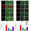
Fig. 3
EXmiR-132-3p infusion rescues synaptic deficits in VD mice. A Representative Golgi staining from cortex and hippocampus regions are shown. Scale bar, 10 μm. B The effects of MSC EX injection on cortical dendritic arborization in Sham and VD mice. CE Representative images and summary data of dendritic spines from cortical and hippocampal neurons from VD mice after treated with EXCon/EXmiR-132-3p/EXantagomiR-132-3p. Data represent the mean ± SEM. n = 20 neurons from 3 mice per group. Three independent experiments were performed. *p < 0.05
Fig. 4
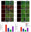
Fig. 4
EXmiR-132-3p infusion reduces cortical neuron loss and abnormal architecture in hippocampal CA1 region in VD mice, injected with various MSC EX. A, C The cortical neuron number in Sham and VD mice was detected by immunofluorescence staining (NeuN, green; DAPI, blue). B, D Representative images and summary data showing the hippocampal neuron number and architecture in VD mice by immunochemistry. Data represent the mean ± SEM. n = 20 fields from 3 mice per group. Three independent experiments were performed. *p < 0.05
Fig. 5
Fig. 5
EXmiR-132-3p infusion reduces cortical neuron apoptosis in VD mice. The apoptotic neurons in cortex were detected by immunofluorescence staining (TUNEL, red; NeuN, green). Scale bar, 30 μm. Data represent the mean ± SEM. n = 20 fields from 3 mice per group. Three independent experiments were performed. *p < 0.05
Fig. 6
Fig. 6
EXmiR-132-3p infusion reduces cortical and hippocampal Aβ production in VD mice. A, C The Aβ production in cortex was detected by immunofluorescence staining (Aβ, red; MAP-2, green). Scale bar, 40 μm. B, D The Aβ production in hippocampus was detected by immunofluorescence staining (Aβ, red; MAP-2, green). Scale bar, 40 μm. Data represent the mean ± SEM. n = 20 fields from 3 mice per group. Three independent experiments were performed. *p < 0.05
Fig. 7
Fig. 7
EXmiR-132-3p infusion reduces cortical and hippocampal Tau hyperphosphorylation in VD mice. A, C The Tau hyperphosphorylation in cortex was detected by immunofluorescence staining (p-Tau, red; MAP-2, green). Scale bar, 40 μm. B, D The Tau hyperphosphorylation in hippocampus was detected by immunofluorescence staining (p-Tau, red; MAP-2, green). Scale bar, 40 μm. Data represent the mean ± SEM. n = 20 fields from 3 mice per group. Three independent experiments were performed. *p < 0.05
Fig. 8
Fig. 8
EXmiR-132-3p incubation increases miR-132-3p level and down-regulated the target protein RASA1 expression in OGD-treated neurons. A Immunofluorescence of MSC EX merged with neurons after incubation (MSC EX, red; β111-tubulin, green). Scale bar, 20 μm. B MiR-132-3p level in OGD-treated neurons was measured by qRT-PCR. C MiR-132-3p regulates the target protein RASA1 expression in OGD-treated neurons. Data represent the mean ± SEM. Three independent experiments were performed. *p < 0.05
Fig. 9
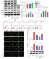
Fig. 9
EXmiR-132-3p incubation decreasing neuron apoptosis and tau phosphorylation by activating Ras/Akt/GSK-3β signaling pathway in OGD-treated neurons. A, B Representative images and summary data showing the level of Ras and phosphorylation of Akt and GSK-3β in OGD-treated neurons after various MSC EX incubation. C The neuron apoptosis was detected by flow cytometry. D, E Representative images and summary data showing the p-Tau level in OGD-treated neurons after various MSC EX incubation (p-Tau, green; nucleus, blue). Scale bar, 40 μm. F The phosphorylation of Tau in OGD-treated neurons after various MSC EX incubation was measured by western blotting. Data represent the mean ± SEM. Three independent experiments were performed. *p < 0.05
Fig. 10
Fig. 10
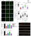
EXmiR-132-3p incubation restores neurite outgrowth and synaptic density in OGD-treated neurons. AC Representative images and summary data showing neurite outgrowth in OGD-treated neurons after various MSC EX incubation (β111-tubulin, green; nucleus, blue). Scale bar, 30 μm. DF Representative images and summary data showing synaptic density in OGD-treated neurons after various MSC EX incubation (PSD95, red; synaptophysin, green). Data represent the mean ± SEM. Three independent experiments were performed. *p < 0.05

Similar articles

Cited by

References

    1. Iadecola C. The pathobiology of vascular dementia. Neuron. 2013;80:844–866. doi: 10.1016/j.neuron.2013.10.008. - DOI - PMC - PubMed
    1. Yang Y, Xu HY, Deng QW, Wu GH, Zeng X, Jin H, et al. Electroacupuncture facilitates the integration of a grafted TrkC-modified mesenchymal stem cell-derived neural network into transected spinal cord in rats via increasing neurotrophin-3. CNS Neurosci Ther. 2021;27:776–791. doi: 10.1111/cns.13638. - DOI - PMC - PubMed
    1. Chen JJ, Zhou SH. Mesenchymal stem cells overexpressing MiR-126 enhance ischemic angiogenesis via the AKT/ERK-related pathway. Cardiol J. 2011;18:675–681. doi: 10.5603/CJ.2011.0032. - DOI - PubMed
    1. Wang L, Qing L, Liu H, Liu N, Qiao J, Cui C, et al. Mesenchymal stromal cells ameliorate oxidative stress-induced islet endothelium apoptosis and functional impairment via Wnt4-beta-catenin signaling. Stem Cell Res Ther. 2017;8:188. doi: 10.1186/s13287-017-0640-0. - DOI - PMC - PubMed
    1. Gu Y, He M, Zhou X, Liu J, Hou N, Bin T, et al. Endogenous IL-6 of mesenchymal stem cell improves behavioral outcome of hypoxic-ischemic brain damage neonatal rats by supressing apoptosis in astrocyte. Sci Rep. 2016;6:18587. doi: 10.1038/srep18587. - DOI - PMC - PubMed

Publication types

MeSH terms

LinkOut - more resources