

Biodistribution of allogenic umbilical cord-derived mesenchymal stromal cells after fetal repair of myelomeningocele in an ovine model
- PMID: 35841029
- PMCID: PMC9284777
- DOI: 10.1186/s13287-022-02991-0
Biodistribution of allogenic umbilical cord-derived mesenchymal stromal cells after fetal repair of myelomeningocele in an ovine model
Abstract
Background: Myelomeningocele (MMC) is a spinal cord congenital defect that leads to paraplegia, sphincter disorders and potential neurocognitive disabilities. Prenatal surgery of MMC provides a significant benefit compared to surgery at birth. Mesenchymal stromal cell (MSC) therapy as an adjuvant treatment for prenatal surgery showed promising results in animal experiments which could be considered for clinical use in human fetuses. Despite numerous reassuring studies on the safety of MSCs administration in humans, no study focused on MSCs biodistribution after a local MSCs graft on the fetal spinal cord.
Aim: The purpose of our study was to assess the biodistribution of umbilical cord-derived mesenchymal stromal cells (UC-MSCs) at birth in lambs who had a prenatal myelomeningocele repair using a fibrin patch seeded with allogenic UC-MSCs.
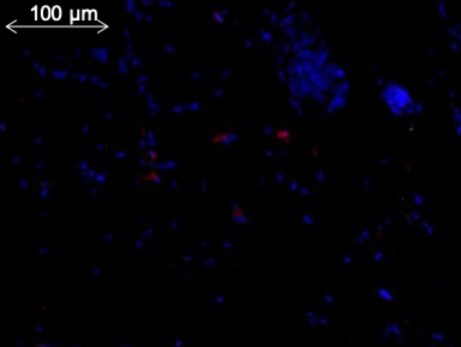
Methods: After isolation, UC-MSCs were tagged using a green fluorescent protein (GFP)-containing lentiviral vector. MMC defects were surgically created at 75 days of gestation and repaired 15 days later using UC-MSCs patch. Lambs were delivered at 142 days and sacrificed. DNA extraction was performed among biopsies of the different organs and q-PCR analysis was used to detect the expression of GFP (GFP DNA coding sequence).
Results: In our 6 surviving lambs grafted with UC-MSCs, GFP lentivirus genomic DNA was not detected in the organs.
Conclusion: These reassuring data will support translational application in humans, especially since the first human clinical trial using mesenchymal stromal cells for in-utero treatment of MMC started recently in U.S.A.
Keywords: Biodistribution; Fetal surgery; Mesenchymal stromal cells; Myelomeningocele; Ovine model; Tracking.
© 2022. The Author(s).
Conflict of interest statement
The authors declare that they have no competing interests.
Figures
References
Publication types
MeSH terms
LinkOut - more resources
Full Text Sources
