. 2022 Jul 15;19(1):187.
doi: 10.1186/s12974-022-02549-0.

Nilotinib modulates LPS-induced cognitive impairment and neuroinflammatory responses by regulating P38/STAT3 signaling

Affiliations

Nilotinib modulates LPS-induced cognitive impairment and neuroinflammatory responses by regulating P38/STAT3 signaling

Jieun Kim et al. J Neuroinflammation. .

Abstract

Background: In chronic myelogenous leukemia, reciprocal translocation between chromosome 9 and chromosome 22 generates a chimeric protein, Bcr-Abl, that leads to hyperactivity of tyrosine kinase-linked signaling transduction. The therapeutic agent nilotinib inhibits Bcr-Abl/DDR1 and can cross the blood-brain barrier, but its potential impact on neuroinflammatory responses and cognitive function has not been studied in detail.

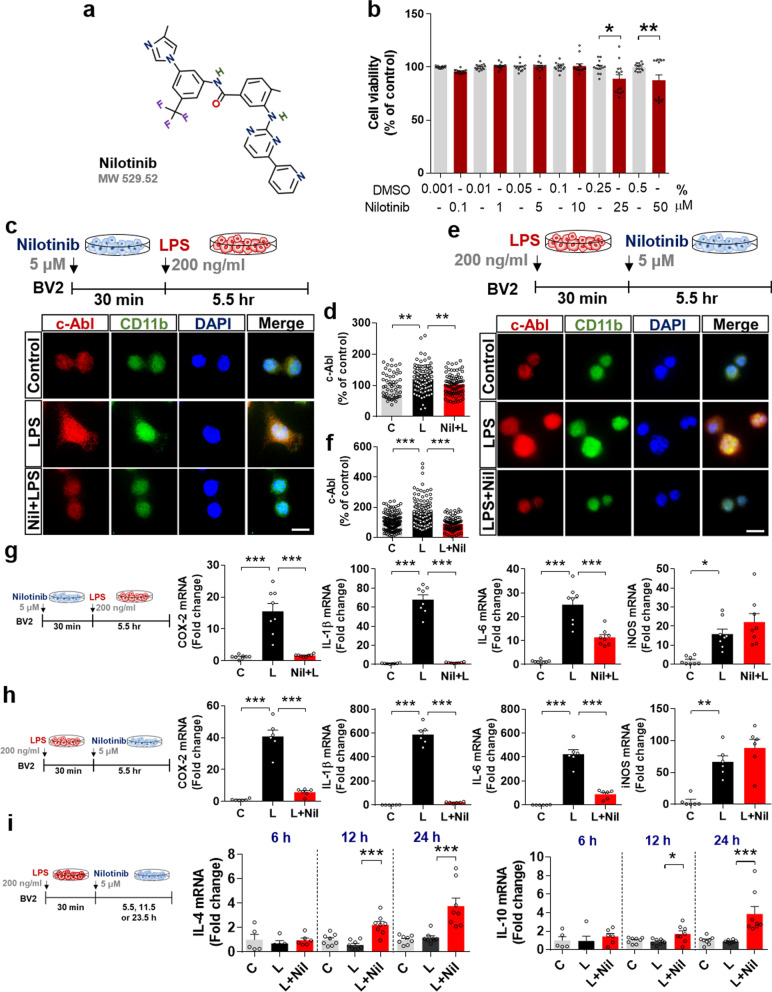
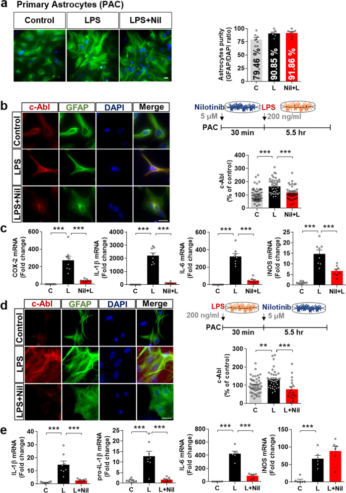
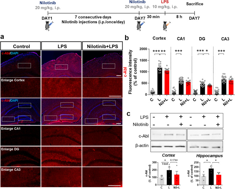
Methods: The effects of nilotinib in vitro and in vivo were assessed by a combination of RT-PCR, real-time PCR, western blotting, ELISA, immunostaining, and/or subcellular fractionation. In the in vitro experiments, the effects of 200 ng/mL LPS or PBS on BV2 microglial cells, primary microglia or primary astrocytes pre- or post-treated with 5 µM nilotinib or vehicle were evaluated. The in vivo experiments involved wild-type mice administered a 7-day course of daily injections with 20 mg/kg nilotinib (i.p.) or vehicle before injection with 10 mg/kg LPS (i.p.) or PBS.

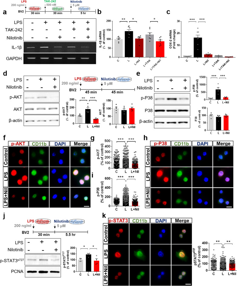
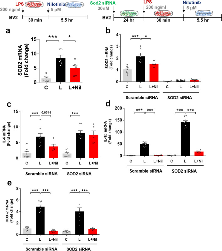
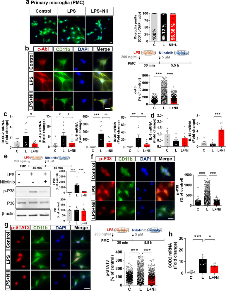
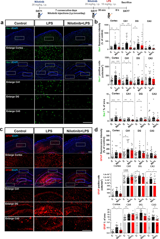
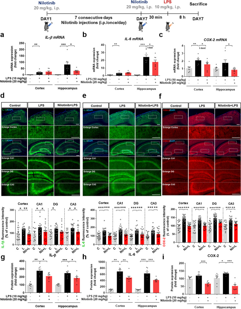
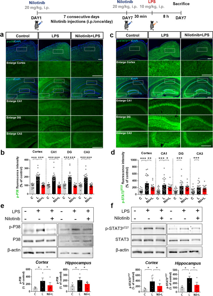
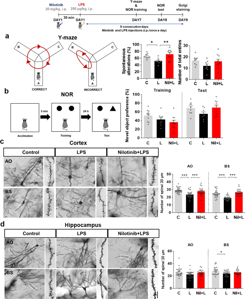
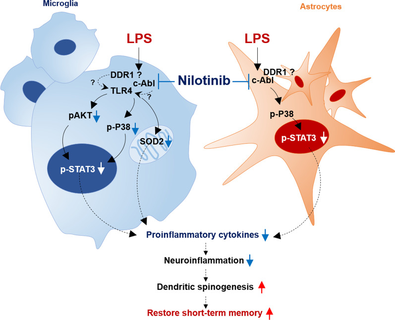
Results: In BV2 microglial cells, pre- and post-treatment with nilotinib altered LPS-induced proinflammatory/anti-inflammatory cytokine mRNA levels by suppressing AKT/P38/SOD2 signaling. Nilotinib treatment also significantly downregulated LPS-stimulated proinflammatory cytokine levels in primary microglia and primary astrocytes by altering P38/STAT3 signaling. Experiments in wild-type mice showed that nilotinib administration affected LPS-mediated microglial/astroglial activation in a brain region-specific manner in vivo. In addition, nilotinib significantly reduced proinflammatory cytokine IL-1β, IL-6 and COX-2 levels and P38/STAT3 signaling in the brain in LPS-treated wild-type mice. Importantly, nilotinib treatment rescued LPS-mediated spatial working memory impairment and cortical dendritic spine number in wild-type mice.

Conclusions: Our results indicate that nilotinib can modulate neuroinflammatory responses and cognitive function in LPS-stimulated wild-type mice.

Keywords: Cognitive function; LPS; Microglia; Nilotinib; SOD2; STAT3; p38.


Conflict of interest statement

The authors have no competing interests to declare.

Figures

Fig. 1
Fig. 1
The multi-tyrosine kinase inhibitor nilotinib suppresses LPS-mediated proinflammatory responses and increases anti-inflammatory responses in BV2 microglial cells. a Structure of nilotinib. b Impact of nilotinib or vehicle on cell viability as assessed by the MTT assay (n = 14/group). c, d Immunocytochemistry of c-Abl expression in LPS-treated BV2 microglial cells pre-treated with nilotinib as shown (C: n = 54; L: n = 141; Nil + L: n = 73). e, f Immunocytochemistry of c-Abl expression in LPS-treated BV2 microglial cells post-treated with nilotinib as shown (C: n = 160; L: n = 130; L + Nil: n = 95). g Real-time PCR analysis of proinflammatory cytokine expression in LPS-treated BV2 microglial cells pre-treated with nilotinib as shown (n = 8/group). h Real-time PCR analysis of proinflammatory cytokine expression in LPS-treated BV2 microglial cells post-treated with nilotinib as shown (n = 6/group). i Real-time PCR analysis of anti-inflammatory cytokine expression in LPS-treated BV2 microglial cells post-treated with nilotinib as shown (n = 5–8/group). C: control, L: LPS, Nil + L: nilotinib + LPS, L + Nil: LPS + nilotinib, *p < 0.05, **p < 0.01, ***p < 0.001, scale bar = 20 μm
Fig. 2
Fig. 2
Nilotinib suppresses LPS-stimulated AKT/P38/STAT3 signaling in BV2 microglial cells. a, b RT-PCR analysis of proinflammatory cytokine IL-1β expression in BV2 microglial cells treated sequentially with LPS, TLR4 inhibitor (TAK-242) and nilotinib as shown (n = 7/group). c Real-time PCR analysis of proinflammatory cytokine COX-2 mRNA expression in BV2 microglial cells treated sequentially with LPS, TLR4 inhibitor (TAK-242) and nilotinib as shown (n = 8/group). d, e Western blotting analysis of AKT/P38 signaling in LPS-treated BV2 microglial cells post-treated with nilotinib as shown (n = 5/group). fi Immunocytochemistry analysis of AKT/P38 signaling in LPS-treated BV2 microglial cells post-treated with nilotinib (p-AKT, C: n = 181; L: n = 162; L + Nil: n = 111, p-P38, C: n = 197; L: n = 246; L + Nil: n = 157). j Western blot analysis of nuclear p-STAT3 expression in LPS-treated BV2 microglial post-treated with nilotinib as shown (n = 4/group). k Immunocytochemistry analysis of p-STAT3 levels in LPS-treated BV2 microglial cells post-treated with nilotinib as shown in j (C: n = 110; L: n = 161; L + Nil: n = 94). C: control, L: LPS, Nil + L: nilotinib + LPS, *p < 0.05, **p < 0.01, ***p < 0.001, scale bar = 20 μm
Fig. 3
Fig. 3
Nilotinib decreases LPS-mediated microglial neuroinflammatory responses in a Sod2-dependent manner. a Real-time PCR analysis of Sod2 gene expression in LPS-treated BV2 microglial cells post-treated with nilotinib as shown (n = 6/group). be Real-time PCR analysis of relative Sod2, IL-6, IL-1β, and COX-2 mRNA levels in BV2 microglial cells transfected with Sod2 siRNA (30 nM) or control (scramble) siRNA for 24 h and subsequently treated with LPS and nilotinib as shown (n = 8/group). C: control, L: LPS, Nil + L: nilotinib + LPS, *p < 0.05, ***p < 0.001
Fig. 4
Fig. 4
Nilotinib alters LPS-evoked changes in pro- and anti-inflammatory cytokine levels via P38/STAT signaling and suppresses Sod2 mRNA levels in primary microglia. a Assessment of primary microglial purity based on the CD11b/DAPI ratio (C: n = 371; L: n = 287; L + Nil: n = 216). b Immunocytochemistry analysis of c-Abl levels in LPS-treated primary microglia post-treated with nilotinib as shown (n = C: n = 561; L: n = 577; Nil + L: n = 298). c, d Real-time PCR analysis of pro- and anti-inflammatory cytokine levels in LPS-treated primary microglia post-treated with nilotinib as shown (n = 8/group). eg Western blotting analysis and immunohistochemistry staining of p-P38 and p-STAT3 levels in LPS-treated primary microglia post-treated with nilotinib as shown (p-P38 for western blot: n = 8/group; p-P38 for ICC: C: n = 526; L: n = 464; Nil + L: n = 352; p-STAT3 for ICC: C: n = 422; L: n = 358; Nil + L: n = 213). h Real-time PCR analysis of Sod2 mRNA in LPS-treated primary microglia post-treated with nilotinib (n = 8/group). *p < 0.05, **p < 0.01, ***p < 0.001. Scale bar = 20 μM
Fig. 5
Fig. 5
Nilotinib downregulates LPS-induced proinflammatory responses in primary astrocytes. a Assessment of primary astrocyte purity based on the GFAP/DAPI ratio (C: n = 253; L: n = 231; L + Nil: n = 236). b Immunocytochemistry analysis of c-Abl expression in LPS-treated primary astrocytes pre-treated with nilotinib as shown (C: n = 59; L: n = 43; Nil + L: n = 42). c Real-time PCR analysis of proinflammatory cytokine levels in LPS-treated primary astrocytes pre-treated with nilotinib as shown (n = 8/group). d Immunocytochemistry analysis of c-Abl expression in LPS-treated primary astrocytes post-treated with nilotinib as shown (C: n = 45; L: n = 39; L + Nil: n = 18). e Real-time PCR analysis of proinflammatory cytokine levels in LPS-treated primary astrocytes post-treated with nilotinib as shown (n = 8/group). C: control, L: LPS, Nil + L: nilotinib + LPS, L + Nil: LPS + nilotinib, **p < 0.01, ***p < 0.001, scale bar = 20 μm
Fig. 6
Fig. 6
Nilotinib suppresses LPS-stimulated P38/STAT3 signaling in primary astrocytes. a, c, e Western blotting analysis of p-P38/p-AKT signaling and p-STAT3/NF-kB transcription factors in LPS-treated primary astrocytes post-treated with nilotinib as shown (n = 5/group). b, d, f, g Immunocytochemistry analysis of p-P38, p-AKT, p-STAT3, and p-NF-kB levels in LPS-treated primary astrocytes post-treated with nilotinib as shown (p-P38, C: n = 17; L: n = 26; L + Nil: n = 15, p-AKT, C: n = 25; L: n = 30; L + Nil: n = 18, p-STAT3, C: n = 126; L: n = 128; L + Nil: n = 173, p-NF-kB, C: n = 203; L: n = 229; L + Nil: n = 213). C: control, L: LPS, L + Nil: LPS + nilotinib, *p < 0.05, **p < 0.01, ***p < 0.001, scale bar = 20 μm
Fig. 7
Fig. 7
Nilotinib reduces LPS-evoked c-Abl levels in the brain in wild-type mice. a Immunofluorescence staining of c-Abl expression in brain slices from wild-type mice injected with vehicle (5% DMSO, 10% PEG and 20% Tween 80 in deionized water) or nilotinib (20 mg/kg, i.p.) daily for 7 days followed by LPS (10 mg/kg, i.p.) or PBS on day 7. b Quantification of the data in a (n = 12–23 brain slices from 3–4 mice/group). c Western blotting analysis of c-Abl protein levels in the cortex and hippocampus (n = 4/group). *p < 0.05, **p < 0.01, ***p < 0.001. Scale bar = 100 μM
Fig. 8
Fig. 8
Nilotinib decreases LPS-induced microglial activation/morphology and astrocyte morphology in the brain in wild-type mice. a, c Immunofluorescence staining of microglial or astrocyte expression in brain slices from wild-type mice injected with vehicle (5% DMSO, 10% PEG and 20% Tween 80 in deionized water) or nilotinib (20 mg/kg, i.p.) daily for 7 days followed by LPS (10 mg/kg, i.p.) or PBS on day 7. b, d Quantification of data in a and c (Iba-1, n = 15–22 brain slices from 3–4 mice/group; GFAP, n = 15–22 brain slices from 3 to 4 mice/group). *p < 0.05, **p < 0.01, ***p < 0.001. Scale bar = 100 μM
Fig. 9
Fig. 9
Nilotinib reduces LPS-mediated IL-1β, IL-6, and COX-2 mRNA and protein levels in wild-type mice. ac Real-time PCR of IL-1β, IL-6, and COX-2 mRNA levels in the cortex and hippocampus (n = 8/group). df Immunofluorescence staining of IL-1β, IL-6, and COX-2 in brain slices from wild-type mice injected with vehicle (5% DMSO, 10% PEG and 20% Tween 80 in deionized water) or nilotinib (20 mg/kg, i.p.) daily for 7 days followed by LPS (10 mg/kg, i.p.) or PBS on day 7. The graphs present the quantification of the raw data from the IF staining in df (IL-1β, n = 14–20 brain slices from 3 to 4 mice/group: IL-6, and COX-2, n = 20–22 brain slices from 3 to 4 mice/group). gi ELISA of IL-1β, IL-6, and COX-2 protein levels in the cortex and hippocampus (n = 8/group). *p < 0.05, **p < 0.01, ***p < 0.001. Scale bar = 100 μM
Fig. 10
Fig. 10
Nilotinib reduces LPS-induced p-P38 and p-STAT3 protein levels in wild-type mice. a, c Immunofluorescence staining of p-P38 and p-STAT3 in brain slices from wild-type mice injected with vehicle (5% DMSO, 10% PEG and 20% Tween 80 in deionized water) or nilotinib (20 mg/kg, i.p.) daily for 7 days followed by LPS (10 mg/kg, i.p.) or PBS on day 7. b, d Quantification of data in a and c (p-P38, n = 14–24 brain slices from 3 to 4 mice/group, p-STAT3, n = 21–24 brain slices from 3 to 4 mice/group). e, f Western blotting analysis of p-P38 and p-STAT3 in the cortex and hippocampus in wild-type mice (n = 4/group). *p < 0.05, **p < 0.01, ***p < 0.001. Scale bar = 100 μM
Fig. 11
Fig. 11
Nilotinib reverses LPS-induced spatial memory impairment and cortical dendritic spine number in wild-type mice. a, b Y-maze and novel object recognition (NOR) tests were performed on days 7 and 8, respectively. Spontaneous alternations and number of total entries are represented in a, and object preference is shown in b (n = 8–11 mice/group). c, d After the behavior experiments, Golgi staining was performed, and dendritic spine number was measured in the cortical and hippocampal AO and BS regions (n = 25–26 neurons from 4 to 5 mice/group). *p < 0.05, **p < 0.01, ***p < 0.001. Scale bar = 10 μM
Fig. 12
Fig. 12
Diagram of the effects of nilotinib on LPS-stimulated neuroinflammatory responses and cognitive function. In microglial cells, nilotinib alters LPS-induced proinflammatory cytokine levels via AKT/P38/STAT3 activation and/or in a Sod2-dependent manner. In primary astrocytes, nilotinib downregulates the LPS-stimulated increase in proinflammatory cytokine levels by inhibiting the P38/STAT3 axis. In wild-type mice, nilotinib reduces LPS-induced gliosis, proinflammatory cytokine levels, and downstream P38/STAT3 signaling. Importantly, nilotinib rescues the short-term memory impairment and decrease in dendritic spinogenesis induced by LPS in wild-type mice. The ability of nilotinib to alter LPS-induced gliosis in vitro/in vivo and to modulate neuroinflammation-linked behavior suggests that nilotinib may have potential utility as a therapeutic drug for neuroinflammation/cognitive-associated disease

References

    1. Pagan FL, Hebron ML, Wilmarth B, Torres-Yaghi Y, Lawler A, Mundel EE, Yusuf N, Starr NJ, Arellano J, Howard HH, et al. Pharmacokinetics and pharmacodynamics of a single dose nilotinib in individuals with Parkinson’s disease. Pharmacol Res Perspect. 2019;7(2):e00470. doi: 10.1002/prp2.470. - DOI - PMC - PubMed
    1. Reinwald M, Schleyer E, Kiewe P, Blau IW, Burmeister T, Pursche S, Neumann M, Notter M, Thiel E, Hofmann WK, et al. Efficacy and pharmacologic data of second-generation tyrosine kinase inhibitor nilotinib in BCR-ABL-positive leukemia patients with central nervous system relapse after allogeneic stem cell transplantation. Biomed Res Int. 2014;2014:637059. doi: 10.1155/2014/637059. - DOI - PMC - PubMed
    1. Manley PW, Cowan-Jacob SW, Mestan J. Advances in the structural biology, design and clinical development of Bcr-Abl kinase inhibitors for the treatment of chronic myeloid leukaemia. Biochim Biophys Acta. 2005;1754(1–2):3–13. doi: 10.1016/j.bbapap.2005.07.040. - DOI - PubMed
    1. Jabbour E, Cortes J, Kantarjian H. Nilotinib for the treatment of chronic myeloid leukemia: an evidence-based review. Core Evid. 2010;4:207–213. - PMC - PubMed
    1. Olivieri A, Manzione L. Dasatinib: a new step in molecular target therapy. Ann Oncol. 2007;18(Suppl 6):vi42–vi46. doi: 10.1093/annonc/mdm223. - DOI - PubMed

MeSH terms