. 2022 Sep;56(5):4653-4668.
doi: 10.1111/ejn.15773. Epub 2022 Jul 22.

Post-fatigue ability to activate muscle is compromised across a wide range of torques during acute hypoxic exposure

Affiliations

Post-fatigue ability to activate muscle is compromised across a wide range of torques during acute hypoxic exposure

Daniel J McKeown et al. Eur J Neurosci. 2022 Sep.

Abstract

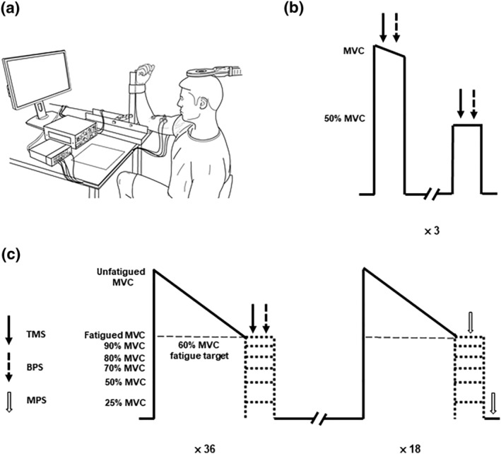
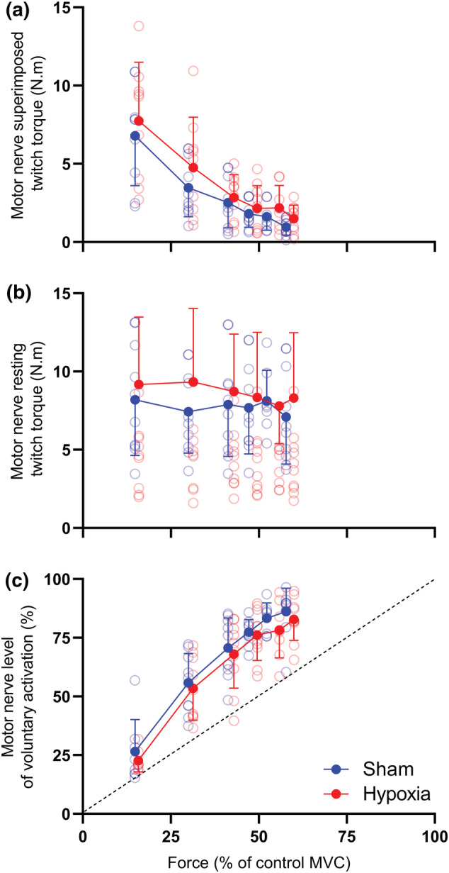
The purpose of this study was to assess how severe acute hypoxia alters the neural mechanisms of muscle activation across a wide range of torque output in a fatigued muscle. Torque and electromyography responses to transcranial and motor nerve stimulation were collected from 10 participants (27 years ± 5 years, 1 female) following repeated performance of a sustained maximal voluntary contraction that reduced torque to 60% of the pre-fatigue peak torque. Contractions were performed after 2 h of hypoxic exposure and during a sham intervention. For hypoxia, peripheral blood oxygen saturation was titrated to 80% over a 15-min period and remained at 80% for 2 h. Maximal voluntary torque, electromyography root mean square, voluntary activation and corticospinal excitability (motor evoked potential area) and inhibition (silent period duration) were then assessed at 100%, 90%, 80%, 70%, 50% and 25% of the target force corresponding to the fatigued maximal voluntary contraction. No hypoxia-related effects were identified for voluntary activation elicited during motor nerve stimulation. However, during measurements elicited at the level of the motor cortex, voluntary activation was reduced at each torque output considered (P = .002, ηp 2 = .829). Hypoxia did not impact the correlative linear relationship between cortical voluntary activation and contraction intensity or the correlative curvilinear relationship between motor nerve voluntary activation and contraction intensity. No other hypoxia-related effects were identified for other neuromuscular variables. Acute severe hypoxia significantly impairs the ability of the motor cortex to voluntarily activate fatigued muscle across a wide range of torque output.

Keywords: corticospinal excitability; exercise; fatigue; hypoxemia; transcranial magnetic stimulation.


Conflict of interest statement

The authors report no conflict of interest.

Figures

FIGURE 1
FIGURE 1
Participant set up (a), control contraction protocol (b) and the fatigue protocol (c). Participants sat in a custom‐built transducer with their right arm secured to measure isometric elbow flexion torque (a). After a 2‐h exposure to hypoxia or sham, the control contraction protocol was performed. The peak maximal voluntary contraction (MVC) torque determined in the control contractions was used to calculate the fatigue target of 60% peak torque (b). Participants performed sets of contractions that each involved a sustained maximal elbow flexion to a fatigue target. Participants immediately received transcranial magnetic (TMS) and brachial plexus stimulations (BPS) or motor nerve stimulations (MNS) at 100%, 90%, 80%, 70%, 50% and 25% of the fatigue target in a randomised order to assess corticospinal and peripheral excitability as well as voluntary activation. Thirty‐six contractions were performed where TMS and BPS stimulation was received, and 18 contractions were performed where MNS stimulation was received, for a total of 54 contractions performed by the participants (c).
FIGURE 2
FIGURE 2
Elbow flexion voluntary torque (a) and biceps brachii and triceps brachii EMGRMS (b) during sham and hypoxia. Elbow flexion torque and EMGRMS data prior to the fatigue protocol (triangles) and following the fatigue protocol (circles). A main effect of contraction intensity was detected for elbow flexion torque (P < .001), biceps brachii EMGRMS (P = .002) and triceps brachii EMGRMS (P < .001). Solid symbols represent the group mean and error bars represent the standard deviation of the mean (n = 10). Individual data are presented as open symbols.
FIGURE 3
FIGURE 3
Post‐fatigue twitch responses to transcranial magnetic stimulation (TMS) and motor nerve stimulation (MNS) during sham and hypoxia. Ensemble average of elbow flexion twitch torque for a single participant are presented for TMS and MNS at 100% (a), 90% (b), 80% (c), 70% (d), 50% (e) and 25% of the fatigued maximal voluntary contraction (MVC). Resting twitch (RT) is also presented for MNS during the sham and hypoxia condition.
FIGURE 4
FIGURE 4
Superimposed twitch torque (a), estimated resting twitch torque (b) and voluntary activation (VA; c) assessed via motor cortical stimulation. (a) Superimposed twitches prior to the fatigue protocol (triangles) and following the fatigue protocol (circles). Twitch responses at 25% maximal voluntary contraction (MVC) torque were not included in calculation of the estimated resting twitch due to the non‐linearity in responses <50% MVC. (b) Although not different prior to fatigue, the estimated resting twitch of the fatigued muscle was significantly lower in hypoxia compared to the sham condition (P = .007). (c) Although not different prior to fatigue, a main effect of condition was detected for VA, where >50% VA measured in hypoxia was lower than VA measured in the sham condition (P = .002; hash represents the condition main effect). Solid symbols represent the group mean and error bars represent the standard deviation of the mean (n = 10). Individual data are presented as open symbols. Dotted line is the line of identity.
FIGURE 5
FIGURE 5
Superimposed twitch torque (a), resting twitch torque (b) and voluntary activation (c) assessed via motor nerve stimulation of the biceps brachii. (a) Superimposed and (b) resting twitches prior to the fatigue protocol (triangles) and following the fatigue protocol (circles). (c) The resultant voluntary activation (VA) was then calculated. Solid symbols represent the group mean and error bars represent the standard deviation of the mean (n = 10). Individual data are presented as open symbols. Dotted line is the line of identity.
FIGURE 6
FIGURE 6
Electromyography (EMG) responses during sham (a) and hypoxia (b) elicited from electrical and magnetic stimulation. Responses from a single participant are presented for electrical stimulation of the brachial plexus (maximal compound action potential, Mmax) and magnetic stimulation of the motor cortex (biceps and triceps motor evoked potentials, MEP) in the fatigued muscle.
FIGURE 7
FIGURE 7
Responses of the corticospinal pathway to motor cortical stimulation. Measures of corticospinal excitability (a), peripheral excitability (b) and corticospinal inhibition (c) prior to the fatigue protocol (triangles) and following the fatigue protocol (circles). Solid symbols represent the group mean and error bars represent the standard deviation of the mean (n = 10).

Similar articles

Cited by

References

    1. Adreani, C. M. , Hill, J. M. , & Kaufman, M. P. (1997). Responses of group III and IV muscle afferents to dynamic exercise. Journal of Applied Physiology, 82(6), 1811–1817. 10.1152/jappl.1997.82.6.1811 - DOI - PubMed
    1. Amann, M. , Blain, G. M. , Proctor, L. T. , Sebranek, J. J. , Pegelow, D. F. , & Dempsey, J. A. (2011). Implications of group III and IV muscle afferents for high‐intensity endurance exercise performance in humans. Journal of Physiology, 589(Pt 21), 5299–5309. 10.1113/jphysiol.2011.213769 - DOI - PMC - PubMed
    1. Amann, M. , Eldridge, M. W. , Lovering, A. T. , Stickland, M. K. , Pegelow, D. F. , & Dempsey, J. A. (2006). Arterial oxygenation influences central motor output and exercise performance via effects on peripheral locomotor muscle fatigue in humans. Journal of Physiology, 575(Pt 3), 937–952. 10.1113/jphysiol.2006.113936 - DOI - PMC - PubMed
    1. Amann, M. , Goodall, S. , Twomey, R. , Subudhi, A. W. , Lovering, A. T. , & Roach, R. C. (2013). AltitudeOmics: On the consequences of high‐altitude acclimatization for the development of fatigue during locomotor exercise in humans. Journal of Applied Physiology, 115(5), 634–642. 10.1152/japplphysiol.00606.2013 - DOI - PMC - PubMed
    1. Amann, M. , Romer, L. M. , Pegelow, D. F. , Jacques, A. J. , Hess, C. J. , & Dempsey, J. A. (2006). Effects of arterial oxygen content on peripheral locomotor muscle fatigue. Journal of Applied Physiology, 101(1), 119–127. 10.1152/japplphysiol.01596.2005 - DOI - PubMed

Publication types