

Assessment of melanoma thickness based on dermoscopy images: an open, web-based, international, diagnostic study
- PMID: 35841304
- PMCID: PMC9796258
- DOI: 10.1111/jdv.18436
Assessment of melanoma thickness based on dermoscopy images: an open, web-based, international, diagnostic study
Abstract
Background: Preoperative assessment of whether a melanoma is invasive or in situ (MIS) is a common task that might have important implications for triage, prognosis and the selection of surgical margins. Several dermoscopic features suggestive of melanoma have been described, but only a few of these are useful in differentiating MIS from invasive melanoma.
Objective: The primary aim of this study was to evaluate how accurately a large number of international readers, individually as well as collectively, were able to discriminate between MIS and invasive melanomas as well as estimate the Breslow thickness of invasive melanomas based on dermoscopy images. The secondary aim was to compare the accuracy of two machine learning convolutional neural networks (CNNs) and the collective reader response.
Methods: We conducted an open, web-based, international, diagnostic reader study using an online platform. The online challenge opened on 10 May 2021 and closed on 19 July 2021 (71 days) and was advertised through several social media channels. The investigation included, 1456 dermoscopy images of melanomas (788 MIS; 474 melanomas ≤1.0 mm and 194 >1.0 mm). A test set comprising 277 MIS and 246 invasive melanomas was used to compare readers and CNNs.
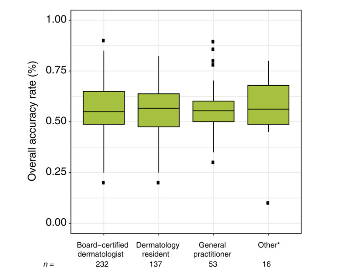
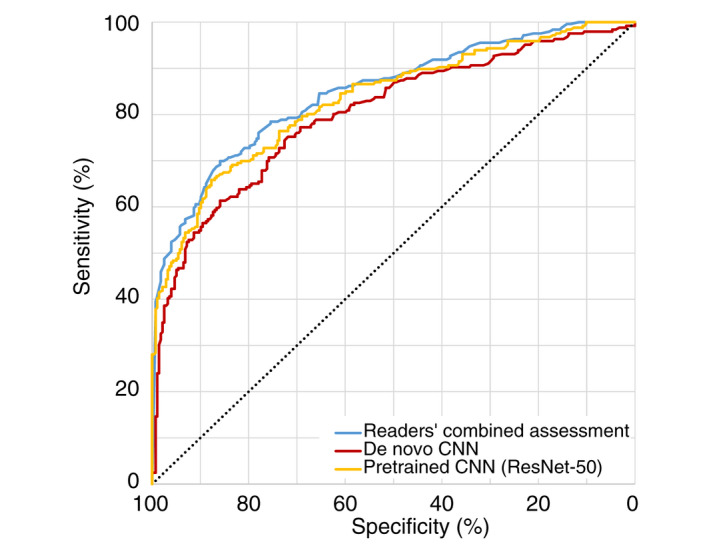
Results: We analysed 22 314 readings by 438 international readers. The overall accuracy (95% confidence interval) for melanoma thickness was 56.4% (55.7%-57.0%), 63.4% (62.5%-64.2%) for MIS and 71.0% (70.3%-72.1%) for invasive melanoma. Readers accurately predicted the thickness in 85.9% (85.4%-86.4%) of melanomas ≤1.0 mm (including MIS) and in 70.8% (69.2%-72.5%) of melanomas >1.0 mm. The reader collective outperformed a de novo CNN but not a pretrained CNN in differentiating MIS from invasive melanoma.
Conclusions: Using dermoscopy images, readers and CNNs predict melanoma thickness with fair to moderate accuracy. Readers most accurately discriminated between thin (≤1.0 mm including MIS) and thick melanomas (>1.0 mm).
© 2022 The Authors. Journal of the European Academy of Dermatology and Venereology published by John Wiley & Sons Ltd on behalf of European Academy of Dermatology and Venereology.
Figures
Similar articles
-
Evaluation of Melanoma Thickness with Clinical Close-up and Dermoscopic Images Using a Convolutional Neural Network.Acta Derm Venereol. 2022 Oct 11;102:adv00790. doi: 10.2340/actadv.v102.2681. Acta Derm Venereol. 2022. PMID: 36172695 Free PMC article.
-
Discrimination Between Invasive and In Situ Melanomas Using Clinical Close-Up Images and a De Novo Convolutional Neural Network.Front Med (Lausanne). 2021 Sep 14;8:723914. doi: 10.3389/fmed.2021.723914. eCollection 2021. Front Med (Lausanne). 2021. PMID: 34595193 Free PMC article.
-
Can Dermoscopy Be Used to Predict if a Melanoma Is In Situ or Invasive?Dermatol Pract Concept. 2021 May 20;11(3):e2021079. doi: 10.5826/dpc.1103a79. eCollection 2021 May. Dermatol Pract Concept. 2021. PMID: 34123569 Free PMC article.
-
Dermoscopy of Nodular Melanoma: Review of the Literature and Report of 3 Cases.Acta Dermatovenerol Croat. 2016 Aug;24(3):203-8. Acta Dermatovenerol Croat. 2016. PMID: 27663921 Review.
-
Enhancing Skin Cancer Diagnosis with Dermoscopy.Dermatol Clin. 2017 Oct;35(4):417-437. doi: 10.1016/j.det.2017.06.003. Epub 2017 Aug 7. Dermatol Clin. 2017. PMID: 28886798 Free PMC article. Review.
Cited by
-
How Does a Convolutional Neural Network Trained to Differentiate between Invasive Melanoma and Melanoma In situ Generalize when Assessing Dysplastic Naevi?Acta Derm Venereol. 2023 Mar 14;103:adv00891. doi: 10.2340/actadv.v103.4822. Acta Derm Venereol. 2023. PMID: 36916955 Free PMC article. No abstract available.
-
Evaluation of Melanoma Thickness with Clinical Close-up and Dermoscopic Images Using a Convolutional Neural Network.Acta Derm Venereol. 2022 Oct 11;102:adv00790. doi: 10.2340/actadv.v102.2681. Acta Derm Venereol. 2022. PMID: 36172695 Free PMC article.
-
Exploring the feasibility of an artificial intelligence based clinical decision support system for cutaneous melanoma detection in primary care - a mixed method study.Scand J Prim Health Care. 2024 Mar;42(1):51-60. doi: 10.1080/02813432.2023.2283190. Epub 2024 Feb 7. Scand J Prim Health Care. 2024. PMID: 37982736 Free PMC article.
-
Reply to Pennington, T.E.; Thompson, J.F. Sentinel Node Biopsy in Melanoma Remains a Valuable Clinical Tool. Comment on "Dixon et al. Primary Cutaneous Melanoma-Management in 2024. J. Clin. Med. 2024, 13, 1607".J Clin Med. 2025 Jan 2;14(1):216. doi: 10.3390/jcm14010216. J Clin Med. 2025. PMID: 39797300 Free PMC article.
-
Performance of a Machine Learning Algorithm on Lesions with a High Preoperative Suspicion of Invasive Melanoma.Acta Derm Venereol. 2024 Jul 18;104:adv40023. doi: 10.2340/actadv.v104.40023. Acta Derm Venereol. 2024. PMID: 39023145 Free PMC article. No abstract available.
References
-
- Vestergaard ME, Macaskill P, Holt PE, Menzies SW. Dermoscopy compared with naked eye examination for the diagnosis of primary melanoma: a meta‐analysis of studies performed in a clinical setting. Br J Dermatol 2008; 159: 669–676. - PubMed
-
- Medical University of Vienna Department of Dermatology . DermaChallenge. 2021. Available at: https://dermachallenge.meduniwien.ac.at [Last accessed October 5, 2021].
MeSH terms
LinkOut - more resources
Full Text Sources
Medical
