. 2023 Feb;280(2):633-641.
doi: 10.1007/s00405-022-07523-3. Epub 2022 Jul 16.

Progressive loss of hearing and balance in superficial siderosis due to occult spinal dural defects

Affiliations

Progressive loss of hearing and balance in superficial siderosis due to occult spinal dural defects

G Michael Halmagyi et al. Eur Arch Otorhinolaryngol. 2023 Feb.

Abstract

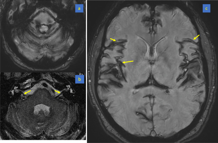
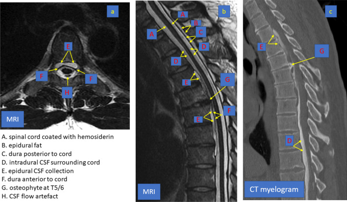
Purpose: Superficial siderosis, a progressive, debilitating, neurological disease, often presents with bilateral impairment of auditory and vestibular function. We highlight that superficial siderosis is often due to a repairable spinal dural defect of the type that can also cause spontaneous intracranial hypotension.

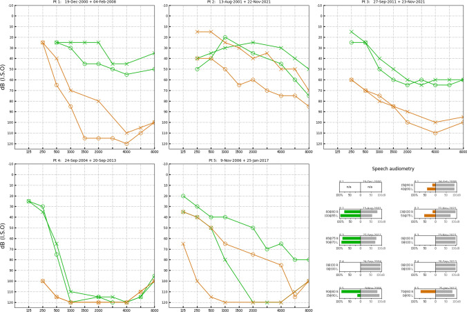
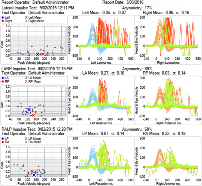
Methods: Retrospective chart review of five patients presenting with moderate to severe, progressive bilateral sensorineural hearing loss as well as vestibular loss. All patients had developed superficial siderosis from spinal dural defects: three after trauma, one after spinal surgery and one from a thoracic discogenic microspur.

Results: The diagnosis was made late in all five patients; despite surgical repair in four, hearing and vestibular loss failed to improve.

Conclusions: In patients presenting with progressive bilateral sensorineural hearing loss, superficial siderosis should be considered as a possible cause. If these patients also have bilateral vestibular loss, cerebellar impairment and anosmia, then the diagnosis is likely and the inevitable disease progress might be halted by finding and repairing the spinal dural defect.

Keywords: CSF leak; Deafness; Spinal dural defects; Superficial siderosis; Vestibulopathy.


Conflict of interest statement

The authors have no conflicts of interest or any financial interests to declare.

Figures

Fig. 1
Fig. 1
Audiograms. The first (shown in green) and last (shown in brown) pure-tone audiogram that we could find for each patient; left ear (x) and right ear (o). In speech audiometry, the stimulus intensity (dB) is shown in grey bars, the comprehension score (%) is shown in green or orange bars, and the result also annotated to the left of each speech test
Fig. 2
Fig. 2
Patient (1). Video head impulse testing showing severe impairment of VOR gain from all 6 semicircular canals. R lateral = 0.06; R anterior = 0.22; R posterior = 0.03; L lateral = 0.05; L anterior = 0.27; L posterior = 0.07. Normal lateral > 0.9; normal vertical > 0.8. Leftward head velocity in blue; rightward head velocity in orange; vestibulo-ocular reflex in green; catch-up saccades in red
Fig. 3
Fig. 3
Patient (4). Susceptibility weighted brain MRI showing extensive leptomeningeal low signal intensity around the cerebellar folia (a), the basis pontis and vestibulocochlear nerves (b) and Sylvian fissures (c) due to hemosiderin deposition
Fig. 4
Fig. 4
Patient (4). T2 weighted spine MRI in axial (a) and sagittal (b) section showing a longitudinally extensive epidural CSF collection from T2 to T 8 (E) with a large spike osteophyte at T5/T6 impinging on the dura (G). The CT myelogram (c) shows contrast in the epidural collection from a CSF leak adjacent to the osteophyte (G). (A) spinal cord coated with hemosiderin. (B) epidural fat—bright on T2. (C) dura posterior to cord; (D) intradural CSF surrounding cord; (E) epidural CSF collection; (F) dura anterior to cord; (G) osteophyte; (H) CSF pulsation artefact
Fig. 5
Fig. 5
Patient (4). Dorsolateral view of spinal cord after laminectomy and opening of dura—rotated up to show the ventral dural defect. A. branch of anterior spinal artery. B. rostral and caudal margins of ventral dural defect with anterior pseudomeningocoele (*) in the depths. C. xanthochromia on spinal cord. D. dentate ligament divided. E. Adhesions to the dural defect, the probable source of chronic subarachnoid hemorrhage

References

    1. Kumar N. Superficial siderosis: a clinical review. Ann Neurol. 2021;89(6):1068–1079. doi: 10.1002/ana.26083. - DOI - PubMed
    1. Parnes SM, Weaver SA. Superficial siderosis of the central nervous system: a neglected cause of sensorineural hearing loss. Otolaryngol Head Neck Surg. 1992;107(1):69–77. doi: 10.1177/019459989210700111. - DOI - PubMed
    1. Pribitkin EA, Rondinella L, Rosenberg SI, Yousem DM. Superficial siderosis of the central nervous system: an underdiagnosed cause of sensorineural hearing loss and ataxia. Am J Otol. 1994;15(3):415–418. - PubMed
    1. Kharytaniuk N, Cowley P, Sayal P, Eleftheriou P, Farmer SF, Chan E, Bamiou DE, Werring DJ (2022) Classical infratentorial superficial siderosis of the central nervous system: pathophysiology, clinical features and management. Pract Neurol. 10.1136/practneurol-2021-003324. - PMC - PubMed
    1. Weekamp HH, Huygen PL, Merx JL, Kremer HP, Cremers CW, Longridge NS. Longitudinal analysis of hearing loss in a case of hemosiderosis of the central nervous system. Otol Neurotol. 2003;24(5):738–742. doi: 10.1097/00129492-200309000-00008. - DOI - PubMed

MeSH terms

LinkOut - more resources